
Page 75 - OR-1-3
P. 75
functional maturation of MSK organoids. For example, Tsigkou et al. seeded collagen-fibronectin gel containing
229
ECs in skeletal muscle not only provide blood supply but HUVECs and human MSCs (hMSCs) on the scaffold.
also regulate the alignment and contractile function of Surprisingly, hMSCs assumed the role of perivascular
muscle fibers by secreting growth factors, such as vascular cells and acted as effective stabilizers of the engineered
endothelial growth factor (VEGF) and insulin-like growth vessels, allowing the HUVECs to form tubular structures
factor (IGF)-1. 223,224 In addition, in the skeletal system, 7 days after scaffold implantation in vivo. Although some
osteogenesis is associated with a specific capillary EC human-derived HUVECs remained after 5 months, the
subtype, termed type H. 218,225 This subtype shows high vascular system of most grafts had been remodeled by
expression of the markers CD31 and endomucin, which are host rat cells. This study confirms the ability of HUVECs
present in the epiphysis and endosteum of postnatal long to function as organoid vascular building cells and to
bones. In addition to inducing vascular growth, type H ECs form connections with blood vessels in vivo to accomplish
also act on osteogenic progenitor cells through molecular nutrient transport. These studies offer a new strategy for
signaling. Therefore, the introduction and integration of inducing angiogenesis in vitro, promoting a significant step
226
a functional vascular network into MSK organoid cultures forward for vascularized organoids, which can effectively
could address the challenge of organoid undernutrition overcome the challenge of oxygen and nutrient diffusion, as
and promote functional maturation of organoids. well as expand the size and scale of organoid culture.
Recently, researchers have made some attempts, 5.1.2. Incomplete innervation
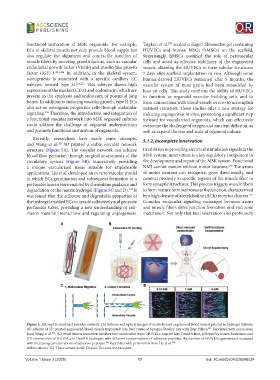
and Wang et al. 227 3D printed a stable vascular network
structure (Figure 5A). The vascular network can achieve In addition to providing electrical stimulation signals in the
blood flow perfusion through surgical anastomosis of the MSK system, innervation is a key regulatory component in
circulatory system (Figure 5B), innovatively providing the development and repair of the MSK system. Functional
230
a unique vascularized tissue suitable for implantable NMJ cannot mature without motor neurons. The axons
applications. Liu et al. developed an in vitro vascular model of motor neurons can recognize, grow directionally, and
in which ECs germination and subsequent formation of a connect precisely to specific regions of the muscle fiber to
perfusable lumen were enabled by chemokine guidance and form synaptic structures. This process triggers muscle fibers
degradation of the matrix hydrogel (Figure 5C and D). It to form mature terminal zones at the junction, characterized
228
was found that the adhesive and degradable properties of by a high density of acetylcholine (ACh) receptor clusters.
231
the hydrogel enabled ECs to invade collectively and generate Complex molecular signaling exchanges between axons
perfusable tubes, providing a new understanding of cell- and muscle fibers drive junction formation and end zone
matrix material interactions and regulating angiogenesis. maturation. Not only that but innervation also profoundly
A B
C D
Figure 5. Attempt to construct vascular network. (A) Scheme and optical images of multichannel engineered blood vessels printed in hydrogel hybrids;
(B) scheme of 3D printed engineered blood vessels implanted into liver tissue of Sprague-Dawley rats with liver failure. Reprinted with permission
227
from Wang et al. ; (C) vessel lumen formation involves two consecutive steps: HUVECs migrate into DexMA first, followed by lumen formation; and
227
(D) intervention of HUVECs in DexMA hydrogels with different concentrations of adhesion peptides, the number of HUVECs germinated increased
with increasing concentrations of adhesion peptides. Reprinted with permission from Liu et al. 228
228
Abbreviations: 3D: Three-dimensional; Dexma: Dextran methacrylate.
Volume 1 Issue 3 (2025) 17 doi: 10.36922/OR025280024