
Page 65 - TD-2-3
P. 65
Tumor Discovery Ureteral IgG4-related disease in urology
fibrosis, and sclerosis of individual glomeruli. The renal
tubule lumens were dilated, accompanied by dystrophic
changes in the tubular epithelium.
Within the lymph node tissue, slight fibrosis was
noted with plasticization of the interfollicular spaces and
sinuses. Morphological changes were most consistent with
tumor-like fibroinflammatory lesions of the ureter, chronic
interstitial nephritis, and sinus histiocytosis of the lymph
nodes.
For the differential diagnosis of IgG4-RD,
immunohistochemistry (IHC) was performed using
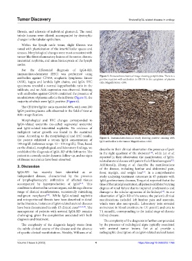
antibodies against CD138, anaplastic lymphoma kinase Figure 5. Immunohistochemical image showing periphlebitis. There is a
positive reaction with antibodies to CD138 in the cytoplasm of plasma
(ALK), kappa and lambda light chains, and IgG4. IHC cells. Magnification: ×200.
specimens revealed a normal kappa/lambda ratio in the
infiltrate, and no ALK expression was observed. Staining
with antibodies against CD138 confirmed the presence of
an admixture of plasma cells in the infiltrate (Figure 5), the
majority of which were IgG4-positive (Figure 6).
The CD138+/IgG4+ ratio exceeded 80%, with over 200
IgG4-positive plasma cells observed in the field of view at
400× magnification.
Morphological and IHC changes corresponded to
IgG4-related ureteritis (so-called segmental ureteritis)
and IgG4-related interstitial nephritis. No evidence of
malignant tumor growth was found in the examined
tissue. According to the morphological and IHC results, Figure 6. Immunohistochemical study showing positive staining with
the patient exhibited a serum IgG4 concentration of IgG4 antibodies in the tumor. Magnification: ×400.
149 mg/dL (reference range: 10 – 135 mg/dL). Thus, based
on the clinical, morphological, and laboratory findings, we describe in their clinical observation the presence of pain
established the diagnosis of IgG4-RD of the left ureter. The in the right quadrant of the abdomen , while Lei et al.
[16]
patient is currently under dynamic follow-up, and no signs reported in their observation the manifestation of IgG4-
of disease recurrence have been observed. related ureteral disease with pain in the left lumbar region .
[12]
3. Discussion Additionally, Zhong et al. describe the manifestations
of the disease, including lumbar and abdominal pain,
IgG4-RD has recently been identified as an fever, myalgia, and weight loss . In a comprehensive
[13]
independent disease, characterized by the presence study analyzing treatment outcomes in 65 patients with
of lymphoplasmacytic infiltration of affected tissues IgG4 genitourinary diseases, Teng et al. reported that at the
accompanied by hyperproduction of IgG4 [1-3] . This time of the initial presentation, all patients exhibited varying
condition is observed in various organs, exhibiting a diverse degrees of renal failure due to impaired urodynamics and
range of clinical manifestations, occasionally mimicking damage to the tubular apparatus of the kidneys . In our
[10]
malignant neoplasms [7,8] . While IgG4-related nephritis observation of IgG4-RD of the ureter, the patient’s clinical
and retroperitoneal fibrosis have been described in detail manifestations included left lumbar pain and anorexia,
in the literature, instances of IgG4-related ureteral diseases which were also non-specific. Laboratory tests revealed
have been documented in only 15 clinical cases [12,13,15] . The an increase in blood creatinine to 131 µmol/L and urea to
management of patients with ureteral IgG4-RD remains 7.1 mmol/L, corresponding to the initial stage of chronic
challenging, given the complexities associated with both kidney disease.
diagnosis and treatment. The complexity of the diagnosis is further compounded
The complexity of the diagnosis largely stems from by the presence of radiological signs typically associated
the subtle clinical course of the disease and the absence with ureteral tumor lesions. Lei et al. provide a
of specific clinical manifestations. Notably, Williams et al. radiographic description of an IgG4-related ureteral lesion
Volume 2 Issue 3 (2023) 4 https://doi.org/10.36922/td.1766