
Page 64 - TD-2-3
P. 64
Tumor Discovery Ureteral IgG4-related disease in urology
A B
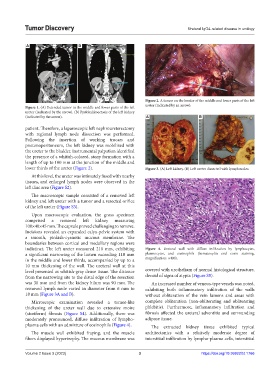
Figure 2. A tumor on the border of the middle and lower parts of the left
ureter (indicated by an arrow).
Figure 1. (A) Extended tumor in the middle and lower parts of the left
ureter (indicated by the arrow). (B) Pyelokalikoectasis of the left kidney
(indicated by the arrow). A B
patient. Therefore, a laparoscopic left nephroureterectomy
with regional lymph node dissection was performed.
Following the insertion of working trocars and
pneumoperitoneum, the left kidney was mobilized with
the ureter to the bladder. Instrumental palpation identified
the presence of a whitish-colored, stony formation with a
length of up to 100 mm at the junction of the middle and
lower thirds of the ureter (Figure 2). Figure 3. (A) Left kidney. (B) Left ureter dissected with lymph nodes.
At this level, the ureter was intimately fused with nearby
tissues, and enlarged lymph nodes were observed in the
left iliac area (Figure S2).
The macroscopic sample consisted of a removed left
kidney and left ureter with a tumor and a resected orifice
of the left ureter (Figure S3).
Upon macroscopic evaluation, the gross specimen
comprised a removed left kidney measuring
100×40×45 mm. The capsule proved challenging to remove.
Incisions revealed an expanded calyx-pelvic system with
a smooth, pinkish-cyanotic mucous membrane. The
boundaries between cortical and medullary regions were
indistinct. The left ureter measured 210 mm, exhibiting Figure 4. Ureteral wall with diffuse infiltration by lymphocytes,
a significant narrowing of the lumen exceeding 110 mm plasmocytes, and eosinophils (hematoxylin end eosin staining,
in the middle and lower thirds, accompanied by up to a magnification: ×400).
10 mm thickening of the wall. The ureteral wall at this
level presented as whitish-gray dense tissue. The distance covered with urothelium of normal histological structure,
from the narrowing site to the distal edge of the resection devoid of signs of atypia (Figure S5).
was 30 mm and from the kidney hilum was 90 mm. The An increased number of venous-type vessels was noted,
removed lymph node varied in diameter from 6 mm to exhibiting both inflammatory infiltration of the walls
18 mm (Figure 3A and B). without obliteration of the vein lumens and areas with
Microscopic examination revealed a tumor-like complete obliteration (non-obliterating and obliterating
thickening of the ureter wall due to extensive moire phlebitis). Furthermore, inflammatory infiltration and
(storiform) fibrosis (Figure S4). Additionally, there was fibrosis affected the ureteral adventitia and surrounding
moderately pronounced, diffuse infiltration of lympho- adipose tissue.
plasma cells with an admixture of eosinophils (Figure 4). The extracted kidney tissue exhibited typical
The muscle wall exhibited fraying, and the muscle architectonics with a relatively moderate degree of
fibers displayed hypertrophy. The mucous membrane was interstitial infiltration by lympho-plasma cells, interstitial
Volume 2 Issue 3 (2023) 3 https://doi.org/10.36922/td.1766