
Page 59 - TD-2-3
P. 59
Tumor Discovery Pleuropulmonary blastoma in child
left-sided chest tube was removed on the 4 post-operative mass (Figure 4B). In the hospital, we worked with the
th
day, while the right-sided chest tube was removed on the medical oncology team to stabilize the patient’s condition
6 day after expansion of the right lung, and no drainage was at the time, but to no avail, he succumbed within 2 days of
th
performed. Post-operative chest X-ray showed complete admission.
expansion of a previously collapsed right lung. A repeat
chest CT scan showed clear bilateral lung zones with no 3. Discussion
evidence of recurrence or metastasis (Figure 4A). The PPB is a rare embryonal thoracic tumor. It is highly
clamshell incision wound was well healed, and the patient aggressive by nature, arising from both lung and pleura, and
was healthy at discharge on the 9 post-operative day. He accounting for 0.5% of all primary malignant lung tumors
th
had been planned for doxorubicin-based chemotherapy in the pediatric population. Three types of PBBs have been
treatment and genetic testing of DICER1, but his parents described in the literature. Type I PBB is purely cystic;
and family members were unwilling to send the patient for type II comprises cystic and solid elements; and type III is
further testing, intervention, and treatment. After 87 days exclusively solid. It is solely confined to the pediatric age
of surgery, he was sent back to the hospital due to severe group, specifically restricted to children under 7 years of
respiratory distress with a recurrence of gross mediastinal age according to most reports . In 2009, a heterozygous
[2]
germline mutation in DICER1 was identified as the first
identified genetic cause in the majority of documented PPB
cases. The DICER1 gene located on human chromosome
14 instructs cells to make a protein called DICER1, which
processes molecules to make microRNA. This germline
mutation in DICER1 can lead to a unique hereditary cancer
predisposition syndrome, which is accompanied by PPB,
cystic nephroma, ovarian Sertoli-Leydig cell tumors, ciliary
body medulloepithelioma, nodular hyperplasia, pituitary
blastoma, pineoblastoma, nasal chondromesenchymal
hamartoma, and differentiated carcinoma of the thyroid
gland. However, a review of 350 confirmed PPB cases
by Messinger et al. confirmed that there were no clinical
differences in terms of germline DICER1 mutation and
thus no distinct prognostic value .
[3]
Type I tumors (median age at diagnosis: 8 months)
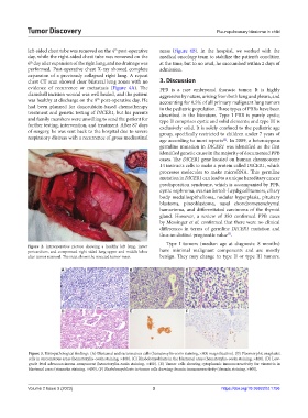
Figure 2. Intraoperative picture showing a healthy left lung, intact
pericardium, and compressed right-sided lung upper and middle lobes have minimal malignant components and are mostly
after tumor removal. The inset shows the resected tumor mass. benign. They may change to type II or type III tumors.
A B C
D E F
Figure 3. Histopathological findings. (A) Blastemal and sarcomatous cells (hematoxylin-eosin staining, ×400 magnification). (B) Pleomorphic anaplastic
cells in sarcomatous areas (hematoxylin-eosin staining, ×400). (C) Rhabdomyoblasts in the blastemal areas (hematoxylin-eosin staining, ×400). (D) Low-
grade fetal adenocarcinoma component (hematoxylin-eosin staining, ×400). (E) Tumor cells showing cytoplasmic immunoreactivity for vimentin in
blastemal areas (vimentin staining, ×400). (F) Rhabdomyoblasts in tumor cells showing desmin immunoreactivity (desmin staining, ×400).
Volume 2 Issue 3 (2023) 3 https://doi.org/10.36922/td.1756