
Page 58 - TD-2-3
P. 58
Tumor Discovery Pleuropulmonary blastoma in child
age of 35 months. Type III PPBs are solid tumors usually like small cells with a high nucleocytoplasmic ratio and a
arising at a median age of 41 months. Type II and type III poorly differentiated tumor. On immunohistochemistry,
tumors are very aggressive and tend to recur or metastasize immunoreactivity for vimentin in blastemal areas with
early. The common sites of metastasis are the brain, liver, focal desmin positivity for rhabdomyoblastic cells was
lymph nodes, pancreas, kidney, and adrenal glands. Type I/ positive but immunonegative for epithelial membrane
Ir PPB has the best prognosis and type III has the least . antigen, S100, and others. A CT scan of the brain was
[2]
The clinical presentations of PPB are non-specific; conducted to rule out metastasis. Screening by abdominal
hence, the chances of misdiagnosis are very high, with a ultrasonography did not yield any unusual findings. The
rd
poor prognosis. Surgery has been cited as the standard patient went into severe hypoxia and shock on the 3 day
treatment, supplemented with chemotherapy for better after admission while undergoing further investigations,
curative effect. Surgical resection is usually challenging and urgent intubation was performed to revive him.
for various reasons, such as the extent and large size of Due to rapid deterioration, surgical intervention was
the tumor, its relation with large vessels and neighboring implemented for the patient after proper consents of
vital structures in the thorax, and neighboring invasion. In his parents were obtained. Under general anesthesia, a
this age group, choosing an incision for proper exposure clamshell incision with bilateral thoracotomy was made.
with maximum lung preservation is also problematic. It is The cystic-solid tumor resulted in the complete collapse of
presumed that surveillance of DICER1 genetic mutation the right lung due to compression. The tumor abutted the
may permit the earlier discovery of PPB, abrogate pericardium and large vessels (both vena cavae and azygos
possible advancement to other types, and improve final vein), and anteriorly crossed the retrosternal space to the
outcomes [2,3] . opposite side. There was no invasion of the pericardial
or left pleural cavity. The left lung was normal. We could
2. Case presentation dissect the tumor almost to its entire extent after gentle
A 5-year-old male with severe respiratory distress coupled dissection and freeing it from large vessels and the anterior
with peripheral desaturation was referred to the emergency surface of the pericardium. After tumor removal with right
department, and he was initially managed with right-sided lower lobe excision, the upper and middle lobes of the right
intercostal tube drain insertion as an emergency measure lung were preserved with complete macroscopic clearance
based on X-ray findings. After temporary stabilization, of the thoracic cavity (Figure 2). The excised tumor was
he was then referred to our institute. Contrast-enhanced around 650 g in weight and approximately 15 × 10 ×
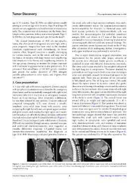
computed tomography (CT) scan showed a sizeable 9 cm in dimensions (Figure 3). The incision was closed in
neoplastic cystic-solid mass lesion with necrotic areas layers with bilateral intercostal drainage tubes. The excised
occupying the right hemithorax, compressing pericardium tumor mass was again sent for detailed histopathological
and great vessels, and causing the right upper and middle examination using immunohistochemical approach. The
lobe bronchus cut-off with the pleural invasion with severe histopathology images showed that tumor has primitive
tracheal and mediastinal shift toward the left side (Figure 1). blastema-like small cells with hyperchromatic nuclei,
Based on CT scan findings of cystic-solid nature of the mass, high nuclear to cytoplasmic ratio, spindled and ovoid
PPB with extraskeletal Ewing’s sarcoma, synovial sarcoma, cells, rhabdomyoblastic cells, and nodules of immature
and embryonal rhabdomyosarcoma were suggested as chondroid elements along with clusters of large anaplastic
possible differential diagnosis. CT-guided biopsy and cells with pleomorphic nuclei. Areas of necrosis were seen
immunohistochemistry established the diagnosis as in the myxoid and fibrous stroma in the tumor (Figure 4).
type II/III PPB. Microscopy indicated primitive blastema- The patient was extubated the 2 day after operation. The
nd
A B C
Figure 1. Computed tomography scan of the chest. (A) Coronal view showing right-sided intercostal tube in tumor mass with gross mediastinal shift.
(B) Sagittal view showing the right hemithorax cystic-solid tumor. (C) Axial view showing right hemithorax cystic-solid tumor, mediastinal shift with
compressed left lung.
Volume 2 Issue 3 (2023) 2 https://doi.org/10.36922/td.1756