
Page 134 - TD-4-1
P. 134
Tumor Discovery An ominous and rare variant of melanoma
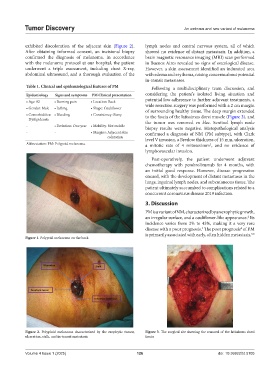
exhibited discoloration of the adjacent skin (Figure 2). lymph nodes and central nervous system, all of which
After obtaining informed consent, an incisional biopsy showed no evidence of distant metastasis. In addition, a
confirmed the diagnosis of melanoma. In accordance brain magnetic resonance imaging (MRI) scan performed
with the melanoma protocol at our hospital, the patient in Buenos Aires revealed no signs of oncological disease.
underwent a triple assessment, including chest X-ray, However, a skin assessment identified an indurated area
abdominal ultrasound, and a thorough evaluation of the with edema and erythema, raising concerns about potential
in-transit metastases.
Table 1. Clinical and epidemiological features of PM Following a multidisciplinary team discussion, and
Epidemiology Signs and symptoms PM Clinical presentation considering the patient’s isolated living situation and
• Age: 82 • Burning pain • Location: Back potential low adherence to further adjuvant treatments, a
• Gender: Male • Itching • Shape: Cauliflower wide resection surgery was performed with a 2 cm margin
of surrounding healthy tissue. The deep margin extended
• Comorbidities: • Bleeding • Consistency: Stony to the fascia of the latissimus dorsi muscle (Figure 3), and
Dyslipidemia
- • Evolution: One year • Mobility: Not mobile the tumor was removed en bloc. Sentinel lymph node
biopsy results were negative. Histopathological analysis
- - • Margins: Adjacent skin confirmed a diagnosis of NM (PM subtype), with Clark
coloration level V invasion, a Breslow thickness of 15 mm, ulceration,
Abbreviation: PM: Polypoid melanoma. a mitotic rate of 4 mitoses/mm , and no evidence of
2
lymphovascular invasion.
Post-operatively, the patient underwent adjuvant
chemotherapy with pembrolizumab for 4 months, with
an initial good response. However, disease progression
ensued, with the development of distant metastases in the
lungs, inguinal lymph nodes, and subcutaneous tissue. The
patient ultimately succumbed to complications related to a
concurrent coronavirus disease 2019 infection.
3. Discussion
PM is a variant of NM, characterized by an exophytic growth,
an irregular surface, and a cauliflower-like appearance. Its
2
incidence varies from 2% to 43%, making it a very rare
disease with a poor prognosis. The poor prognosis of PM
4
3
is primarily associated with early, often hidden metastasis,
5,6
Figure 1. Polypoid melanoma on the back
Figure 2. Polyploid melanoma characterized by the exophytic tumor, Figure 3. The surgical site showing the removal of the latissimus dorsi
ulceration, stalk, and in-transit metastasis fascia
Volume 4 Issue 1 (2025) 126 doi: 10.36922/td.5105