
Page 101 - AN-2-2
P. 101
Advanced Neurology Inflammatory myopathies during COVID-19
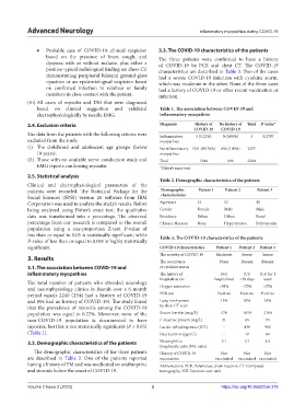
• Probable case of COVID-19: clinical suspicion 3.3. The COVID-19 characteristics of the patients
based on the presence of fever, cough, and The three patients were confirmed to have a history
dyspnea with or without malaise, plus either a of COVID-19 by PCR and chest CT. The COVID-19
positive typical radiological finding on chest CT characteristics are described in Table 3. Two of the cases
demonstrating peripheral bilateral ground-glass had a severe COVID-19 infection with cytokine storm,
opacities or an epidemiological suspicion based which was moderate in the other. None of the three cases
on confirmed infection in relatives or family had a history of COVID-19 or other recent vaccination or
members in close contact with the patient. infection.
(iii) All cases of myositis and DM that were diagnosed
based on clinical suggestion and validated Table 1. The association between COVID‑19 and
electrophysiologically by needle EMG. inflammatory myopathies
2.4. Exclusion criteria Diagnosis History of No history of Total P‑value*
COVID‑19 COVID‑19
The data from the patients with the following criteria were Inflammatory 3 (0.22%) 0 (0.00%) 3 0.2797
excluded from the study: myopathies
(i) The childhood and adolescent age groups (below No inflammatory 1341 (99.78%) 896 (100%) 2237
18 years). myopathies
(ii) Those with no available nerve conduction study and Total 1344 896 2240
EMG reports confirming myositis. *Fisher’s exact test.
2.5. Statistical analysis
Table 2. Demographic characteristics of the patients
Clinical and electrophysiological parameters of the
patients were recorded. The Statistical Package for the Demographic Patient 1 Patient 2 Patient 3
Social Sciences (SPSS) version 26 software from IBM characteristics
Corporation was used to analyze the study’s results. Before Age/years 34 53 40
being analyzed using Fisher’s exact test, the qualitative Gender Female Male Male
data was transformed into a percentage. The observed Residency Urban Urban Rural
percentage from our research is compared to the overall Chronic illnesses None Hypertension Polymyositis
population using a one-proportion Z-test. P-value of
less than or equal to 0.05 is statistically significant, while Table 3. The COVID‑19 characteristics of the patients
P-value of less than or equal to 0.001 is highly statistically
significant. COVID‑19 characteristics Patient 1 Patient 2 Patient 3
The severity of COVID-19 Moderate Severe Severe
3. Results
The occurrence None Present Present
3.1. The association between COVID-19 and of cytokine storm
inflammatory myopathies The history of Not ICU ICU for 1
hospitalization hospitalized >30 days week
The total number of patients who attended neurology
and neurophysiology clinics in Basrah over a 6-month Oxygen saturation >93% <70% <70%
period equals 2240 (2344 had a history of COVID-19 PCR test Positive Positive Positive
and 896 had no history of COVID-19). The study found Lung involvement 15% 50% 55%
that the prevalence of myositis among the COVID-19 by chest CT scan
population was equal to 0.22%. Moreover, none of the Serum ferritin (mcg/L) 670 1650 2100
non-COVID-19 population is documented to have C-reactive protein (mg/L) 21 66 95
myositis, but this is not statistically significant (P > 0.05) Lactate dehydrogenase (U/L) - 430 980
(Table 1). Interleukin-6 (pg/mL) - 19 88
3.2. Demographic characteristics of the patients Neutrophil to 3.1 3.7 4.9
lymphocyte ratio (N/L ratio)
The demographic characteristics of the three patients History of COVID-19 Not Not Not
are described in Table 2. One of the patients reported vaccination vaccinated vaccinated vaccinated
having a history of PM and was medicated on azathioprine Abbreviations: PCR: Polymerase chain reaction; CT: Computed
and steroids before the onset of COVID-19. tomography; ICR: Intensive care unit.
Volume 2 Issue 2 (2023) 3 https://doi.org/10.36922/an.378