
Page 104 - AN-2-2
P. 104
Advanced Neurology Inflammatory myopathies during COVID-19
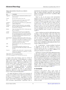
Table 6. Characteristics of myositis cases related to neuroinvasion; (b) neurological complications developing
COVID‑19 after the initial infectious symptoms and supporting
indirect mechanisms (“postinfectious” hypothesis), likely
Item Description immune-mediated .
[6]
Diagnosis Six confirmed cases of polymyositis and four cases of
dermatomycosis Based on the in vivo and in vitro neuroinvasive
Gender Six were women, and four were men capacities of SARS-CoV and MERS-CoV, with which the
Age The minimum and maximum ages were 23 and 77, etiological agent of COVID-19 (i.e., SARS-CoV-2) shares
respectively, with a mean age of 55.6 years 79.5% and 50% gene homology, respectively, the capacity
COVID-19 Nine patients had positive PCR and one patient had a of SARS-CoV-2 to invade the nervous system has been
[23]
history positive serological test hypothesized . Given the early onset of anosmia and
COVID-19 Critical in one case, severe in four cases, moderate ageusia, one idea is that olfactory, trigeminal, or gustative
severity in one case, mild in two cases, and in two cases, the terminals may serve as entry points for the virus, which
severities were not reported may then propagate to the central nervous system (CNS)
Relapse Three cases relapsed after COVID-19 and seven cases through retrograde axonal transport and trans-synaptic
got a new diagnosis transfer .
[24]
Clinical All patients presented with lower and upper limb Lower cranial nerves might be additional entry routes,
features proximal muscle weakness; three reported myalgia;
two reported dysphagia; three reported a typical rash; resulting in early involvement of the lower brain stem
one reported respiratory muscle weakness; and one and perhaps explaining some specific characteristics of
had cardiac involvement COVID-19, such as hypoxia out of proportion to dyspnea
[25]
Immunological Anti-nuclear antibodies were positive in three cases, and the frequency of syncope . Alternative methods
tests autoantibodies were negative in five cases. Anti-MI 2b of neuroinvasion applicable to both the CNS and PNS
was positive in one patient; anti-Ro/SSA antibodies include entrance by circulating immune cells, infection
were positive in one patient; and an anti-Smith
antibody was positive in one patient of the vascular endothelium, and passing the blood-brain
[6]
CPK Markedly elevated in nine cases, and three reported barrier or blood-nerve barrier .
mild elevation The “postinfectious” immune-mediated hypothesis
EMG Two patients had myopathic potential, one was is supported by the fact that COVID-19 induces a
normal, and for the other seven patients, an EMG was proinflammatory state due to the production of several
not done cytokines, including IL1, IL6, and TNF, in addition to the
Outcome One patient died, and the other 11 improved on either subsequent activation of immune cells .
[24]
steroids alone (4 patients) or a combination of steroids
and immunomodulators Furthermore, the gene-level biology of this phenomenon
[26]
Abbreviations: CPK: Creatine phosphokinase; is still poorly understood , despite the extensive study
EMG: Electromyography; PCR: Polymerase chain reaction. conducted in the SARS-CoV-2 area. In addition, the viral
infection of endothelial cells leads to the breakdown of the
it triggers. Early tissue damage caused by COVID-19 blood–brain barrier, which induces acute inflammation and
increases the production of proinflammatory cytokines, causes neuronal injury and disruption of neurogenesis. In
which attract more inflammatory cells and increase addition, these proinflammatory cytokines may influence
inflammatory reactants . Repeat and amplification of this vascular remodeling, resulting in the loss of vascular wall
[21]
process result in what is often referred to as a “cytokine integrity, which may lead to intraparenchymal bleeding.
storm” or a systemic inflammatory response described Finally, cytokine storming is an established risk factor for
as macrophage activation syndrome or secondary coagulopathy, which may trigger a stroke [27,28] .
hemophagocytic lymphohistiocytosis . COVID-19’s
[22]
potential to predispose the body to immunological 5. Conclusion
hyperactivity allows us to predict the development of post- COVID-19 could present with a wide variety of
COVID-related autoimmune disorders .
[10]
neurological complications, including PM and DM, and
The causal link between COVID-19 and symptoms of the prevalence of these disorders increased noticeably after
nervous system problems has been determined purely based the pandemic. It is important to highlight such disorders
on their co-occurrence in time. Two patterns have been and increase awareness about them as probable sequelae
documented: (a) neurological complications occurring of COVID-19 in order not to misdiagnose them, which
together with COVID‐19 symptoms and suggesting a direct might lead to serious complications, including respiratory
viral mechanism (“parainfectious” hypothesis), such as failure and death.
Volume 2 Issue 2 (2023) 6 https://doi.org/10.36922/an.378