
Page 70 - AN-2-4
P. 70
Advanced Neurology A novel marker for healthy intracranial arteries
Table 3. Univariate and multivariate analysis of the FB sign
Variable Univariate analysis Multivariate analysis
Presence of FB Absent or abnormal P‑value OR (95% CI) P‑value
sign (n=381) FB sign (n=624)
Moderate ICA stenosis (n [%]) 96 (25.1) 208 (33.4) <0.001* 0.39 (0.28 – 0.54) <0.001*
Severe ICA stenosis (n [%]) 24 (6.3) 221 (35.5) 0.10 (0.06 – 0.17) <0.001*
MCA Stenosis degree (median % [IQR]) 0 (0, 23) 20 (0, 49) 0.010* 0.85 (0.80 – 0.90) <0.001*
Bifurcation angle, (x±SD degree [°]) 96±18 102±22 <0.001* 0.86 (0.79 – 0.93) <0.001*
Note: *P<0.05. Abbreviations: CI: Confidence interval; FB: Fried-breadstick; ICA: Internal carotid artery; IQR: Interquartile range; MCA: Middle
cerebral artery; OR: Odds ratio; SD: Standard deviation.
A patients with intracranial atherosclerosis in future trials,
with no additional costs or risks.
The underlying mechanisms of the FB sign are intricate
and remain unclear. TOF-MRA is a gradient echo sequence
based on the flow-related enhancement theory . The
[16]
signal intensity of the intracranial arteries on TOF-MRA
is associated with flow velocities in the vessel lumen.
Both high velocity and low velocity of blood flow, or
B in-plane blood flow, could cause signal loss [24,25] . Saloner
et al. investigated central signal loss with computational
[26]
fluid dynamics, phantom, and healthy volunteers. They
simulated parabolic laminar flow and demonstrated that
slow peripheral laminar flow formed a counter-rotating
secondary flow when entering a curved vessel, leading to
central intraluminal signal loss from saturation of the spin
magnetization . The study supported the idea that the
[26]
combination of parabolic laminar flow and ICA curvature
contributed to the signal loss, which matched our
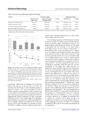
Figure 3. The prevalence of the Fried-Breadstick sign in different groups observation. Since other flow types were not simulated in
and MCA stenosis. (A) The FB sign is most frequently present in plaque-
free MCAs and least frequently in MCAs with non-SSI. *P < 0.005, the above-mentioned study, another possible explanation
**P < 0.001. (B) The FB sign prevalence decreases as the MCA stenosis for the central signal loss is the spiral laminar flow. Spiral
degree increases. To note, the MCA stenosis is measured in terms of laminar flow demonstrates a relatively low velocity or
area stenosis (refer to Figure S2) for the prevalence change of the Fried- in-plane blood flow in the rotational axis and high velocity
Breadstick sign concerning MCA diameter stenosis. The error bar around the rotational axis . The spiral laminar flow is
[5]
represents the 95% confidence interval.
Abbreviations: FB: Fried-breadstick; MCA: Middle cerebral artery; created by the rotational compressive pumping of the
SSI: Single subcortical infarct. heart and maintained by the multi-planar tapered, curved,
and branching arterial geometry [5,27] . Mostly seen in large
vasculatures. Additionally, it investigated the relationship arteries, including ICAs [6,28] , the spiral flow contributes to
between the presence of the FB sign and ICAD. We the relatively uniform wall shear stress in the bifurcation
[29]
observed that the FB sign is more frequently present in region , flow stabilization, and the suppression of flow
[30]
plaque-free MCAs or in cases of low-grade ICA-MCA disturbance and stagnation . The loss of spiral laminar
stenosis compared to high-grade ICA-MCA stenosis. Even flow has been reported to be associated with the presence
[27]
in atherosclerotic MCAs without luminal narrowing, the FB and progression of atherosclerotic diseases . Although
sign was less frequently presented than in plaque-free MCAs, turbulent flow was not simulated in curved vasculature
suggesting the absent or abnormal FB sign is associated in previous literature, it is unlikely for turbulent flow
with atherosclerosis. These results support our hypothesis to manifest as a uniform central signal loss, given its
that the FB sign is associated with a relatively healthy ICA- complicated flow direction and speed. Turbulent flow can
MCA vasculature without severe atherosclerosis. Since the lead to dephasing artifacts in TOF-MRA [31,32] , manifested
FB sign can be easily identified and assessed on routine as signal loss affecting both the central and peripheral of
TOF-MRA, it holds promise for stratifying stroke risk in the vessel lumen.
Volume 2 Issue 4 (2023) 6 https://doi.org/10.36922/an.1238