
Page 88 - AN-2-4
P. 88
Advanced Neurology Narcolepsy-multiple sclerosis coexistence
and Hoffman signs, a positive Babinski sign on the right, a At the beginning, the patient received a course of
positive Romberg sign, and an ataxic gait. shock therapy consisting of 1.0 g methylprednisolone
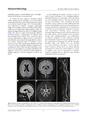
As revealed by brain magnetic resonance imaging administered daily for a consecutive 3 days. The dosage
(MRI), multiple lesions with high T2 or FLAIR signals was then gradually tapered to 500 mg, 250 mg, and
were present in the deep cerebral white matter surrounding 120 mg, each lasting 3 days. Altogether, the entire
the two lateral ventricles (Figure 2A-C). No abnormalities treatment course accounted for 12 days. Subsequently, the
were observed in magnetic resonance angiography patient was discharged with 60 mg oral prednisone and
(Figure 2D). In addition, whole spine MRI showed multiple received subcutaneous administration of ofatumumab.
small punctate hyperintense signal shadows within the In addition, various symptomatic management measures
medulla oblongata and spinal cord on T2-weighted images were implemented, including gastric protection,
(Figure 2E). Data on visual evoked potentials indicated potassium supplementation, prevention of osteoporosis,
less-than-satisfactory differentiation of bilateral P100 and improvement of limb numbness. After synergistic
and prolonged latency. Results from cerebrospinal fluid therapy with methylprednisolone and ofatumumab,
puncture demonstrated a slight elevation in white blood the patient’s symptoms of limb numbness, unsteady
cells, lgG level, and protein level, along with the presence gait, and daytime sleepiness improved significantly. On
of a positive oligoclonal band. To establish a clearer the manifestation of symptoms, the patient’s expanded
distinction from other demyelinating disorders affecting disability status scale (EDSS) score was recorded
the nervous system, the patient underwent supplementary as 2 points. Follow-up was done 1 month after the
examinations to detect MOG and AQP-4 antibodies, which initial treatment, during which the patient achieved
turned out to be negative. In accordance with McDonald’s improvement in EDSS score, which reduced to 1 point.
diagnostic criteria, these clinical and ancillary diagnostic In addition, there was an improvement in daytime
findings supported a definitive diagnosis of MS in this sleepiness symptoms and in nighttime sleep disorders
case . compared to before the treatment.
[4]
A B E
C D
Figure 2. Magnetic resonance imaging (MRI) on July 14, 2023. (A-C) Axial T2-FLAIR sequences of cranial MRI show multiple hyperintense periventricular
lesions around the lateral ventricles, as indicated by arrows. (D) Magnetic resonance angiography reveals no significant abnormalities. (E) T2-weighted
sequences of whole spine MRI demonstrate multiple hyperintense lesions within the medulla oblongata and spinal cord, as indicated by arrows.
Volume 2 Issue 4 (2023) 3 https://doi.org/10.36922/an.1913