
Page 13 - ARNM-2-4
P. 13
Advances in Radiotherapy
& Nuclear Medicine Radionuclide-carrying liposomes
6. Diagnostic applications of radionuclide- cells encounter when they metastasize from a cancerous
containing liposomes site in the body. Localizing the sentinel lymph node and
removing it for pathological examination is important.
Liposomes labeled with the most commonly used single
photon agent, 99m Tc, were shown by Goins et al. to be A novel method of trapping liposomes in sentinel
26
useful for detecting sites of cancer, sites of infection, and lymph nodes was developed by Zavaleta et al. In this
99m
inflammation. 22,23 Investigators from the Netherlands have method, Tc-radiolabeled liposomes were coated with
also shown the potential application of 99m Tc liposomes biotin and simultaneously injected with avidin around a
tumor. Their results showed both the biotin and avidin
in detecting sites of infection and inflammation. aggregating as they moved toward the sentinel lymph
24
Liposomes accumulate at sites of infection in the same node and became entrapped. In this example, the 99m Tc-
manner they accumulate in tumors following intravenous liposomes also had encapsulated blue dye to guide the
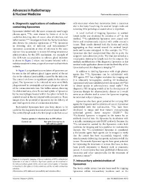
administration, by the EPR mechanism. An example of surgeon’s radio-detection probe. The blue dye served as a
this EPR-type accumulation in salivary gland infection visual guide, staining the lymph node for the surgeon. The
is shown in Figure 2 where rats became infected with a multiple modifications of the diagnostic liposomes in this
sialodacryoadenitis virus, a type of coronavirus that infects sentinel node application demonstrate the flexibility of the
rodents. liposomal system for diagnostic imaging. 25,26
In Figure 2, a significant accumulation of liposomes can In addition to labeling liposomes with single photon
be seen in the left salivary gland (upper arrow) of the rat agents like 99m Tc, liposomes can be radiolabeled with
due to the enhanced permeability caused by the infection. PET agents. PET has a higher resolution for imaging and
The control rat shows no significant uptake in the salivary it is inherently tomographic, enabling 3D localization.
gland. The liposomes in the infected rat were most likely Liposomes have been radiolabeled with zirconium-89 ( Zr)
89
phagocytosed by neutrophils and macrophages at the site to detect uptake in atherosclerotic vessel walls. This
27
of the coronavirus infection. The hollow arrows, denoting diagnostic PET imaging would aid in the development of
the abdominal area, show the normal uptake of liposomes liposome therapy for atherosclerotic disease as it would
by the macrophages located within the spleen in both the serve as an effective tool to screen for liposome targeting
control rat and in the rat infected with coronavirus. These in individual subjects’ plaques.
images show the potential of liposomes for the delivery of Liposomes also have great potential for carrying PET
anti-viral agents to sites of coronavirus infection. agents for diagnosis and localization of cancer. Liposomes
Radiolabeled liposomes have also been shown to be radiolabeled with the radionuclide, 64 Cu, have been
useful for the diagnostic location of sentinel lymph nodes. developed to image and treat tumors overexpressing
25
28
The sentinel lymph node is the first lymph node that cancer epidermal growth factor receptor (EGRF). The
64 Cu-labeled liposome is targeted to the tumor by an
antibody inserted into the liposome by incubation with
a micelle containing an anti-EGRF antibody. Because the
64 Cu-radionuclide carried by the liposomes emits both
positrons for imaging and beta particles for radionuclide
therapy, this Cu-radiolabeled liposome is considered
64
a theranostic agent. Such agents can be imaged to verify
that the cancer has been successfully targeted, while
simultaneously delivering therapeutic radiation to kill the
tumor. Theranostic liposomal agents have great potential
for improving the treatment of tumors that have, to date,
been poorly responsive to therapy such as pancreatic
cancer. A recent review has been published of theranostic
liposomes and other nanoparticles for the treatment of
pancreatic cancer. 28
Figure 2. Scintigraphic imaging of the accumulation of 99 Tc-radiolabeled
m
liposomes in an area of infection. Significant uptake can be visualized in 7. Intracavitary administration of
the infected salivary gland (white arrow) and normal uptake of liposome liposomes
is visualized in the spleen in both control rat and the rat with coronavirus
infection. The biotin-avidin system has also been used for trapping
Abbreviation: EPR: Enhanced permeability and retention effect. liposomes within intracavitary locations such as the
Volume 2 Issue 4 (2024) 5 doi: 10.36922/arnm.4373