
Page 89 - BH-2-2
P. 89
Brain & Heart Left PAPVC to left atrial appendage anastomosis
that is, anastomosis connecting vertical vein to left atrial course was confirmed. The junction of the vertical vein
appendage, and highlight the procedural nuances for to the innominate vein was then divided and transfixed
patients with distinct age difference. (Figure 2B). Left atrial appendage was opened longitudinally
on its dorsal surface after inducing cardioplegic cardiac
2. Case presentation arrest. The open end of the vertical vein was fashioned to
2.1. Case 1 facilitate its anastomosis to the left atrial appendage opening
without any twist or tension, using a 5-0 polypropylene
The first case is a 32-year-old patient presented with continuous suture (Figures 2C-E, and 3). The ASD was
symptoms of intermittent palpitations and breathlessness closed using autologous pericardium (Figure 2F).
for a couple of years. According to an echocardiography
study, the patient had a large secundum ASD with a left Intraoperative transesophageal echocardiography
upper pulmonary vein and lingual vein draining through studies conducted in different time sessions, that is,
a vertical vein to an innominate vein. All right-sided immediately after the surgery and following a 6-month
pulmonary veins and the left lower vein were draining follow-up, revealed no gradient in the left atrial appendage
normally, confirmed by the catheterization study (Figure 1). anastomosis (Figure 4).
The patient was surgically treated with routine median 2.2. Case 2
sternotomy and cardiopulmonary bypass, which involved
dissecting the vertical vein and looping it in the left The second case is a 15-day-old neonate who developed
paracardiac gutter (Figure 2A). All the pulmonary veins respiratory distress and bluish discoloration of extremities
joining the vertical vein were dissected and their anatomical since birth. An echocardiography study revealed that
the neonatal patient was diagnosed with the mixed-
A B type TAPVC with restrictive ASD, characterized by the right
pulmonary veins directly draining into the coronary sinus,
and the left pulmonary veins draining to the innominate
vein through a left-sided vertical vein (Figure 5A).
Due to obstructed atrial communication, median
sternotomy was implemented, and cardiopulmonary bypass
was established with an aortobicaval cannulation. The
vertical vein was dissected in the left paracardiac gutter after
cardioplegic cardiac arrest was induced. All the pulmonary
veins joining the vertical vein were dissected around the
vertical vein. The vertical vein was then ligated and clipped
Figure 1. Catheterization study images. (A) Left inferior pulmonary vein
draining normally to the left atrium. (B) Left superior pulmonary vein at the innominate vein junction without dividing it and
and lingual vein draining to innominate vein (IV) via vertical vein (VV). proximally controlled with silk suture loop (Figure 5B).
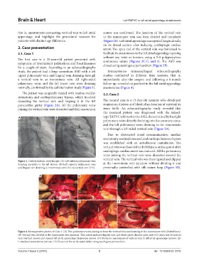
A B C
D E F
Figure 2. Intraoperative photos of Case 1. (A) Two pulmonary veins joining to form the vertical vein and draining to the innominate vein (dotted lines).
(B) Vertical vein divided at the innominate vein junction. Thin arrow indicates ligated end, and thick arrow denotes open end. (C) Open end of vertical
vein (vertical arrow) and opened left atrial appendage (transverse arrow). (D) Posterior anastomosis of vertical vein to left atrial appendage (arrow). (E)
Completed anastomosis (arrow). (F) Closure of the atrial septal defect using autologous pericardium.
Volume 2 Issue 2 (2024) 2 doi: 10.36922/bh.2376