
Page 85 - BH-2-2
P. 85
Brain & Heart Case of rhombencephalitis and hydrocephalus
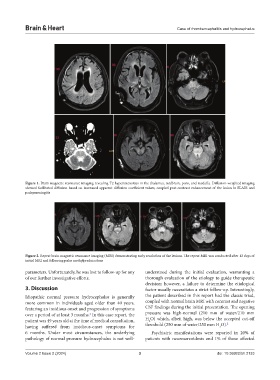
Figure 1. Brain magnetic resonance imaging revealing T2 hyperintensities in the thalamus, midbrain, pons, and medulla. Diffusion-weighted imaging
showed facilitated diffusion based on increased apparent diffusion coefficient values, coupled post-contrast enhancement of the lesion in FLAIR and
pachymeningitis
Figure 2. Repeat brain magnetic resonance imaging (MRI) demonstrating early resolution of the lesions. The repeat MRI was conducted after 12 days of
initial MRI and following pulse methylprednisolone
parameters. Unfortunately, he was lost to follow-up for any understood during the initial evaluation, warranting a
of our further investigative efforts. thorough evaluation of the etiology to guide therapeutic
decision; however, a failure to determine the etiological
3. Discussion factor usually necessitates a strict follow-up. Interestingly,
Idiopathic normal pressure hydrocephalus is generally the patient described in this report had the classic triad,
more common in individuals aged older than 40 years, coupled with normal brain MRI with contrast and negative
featuring an insidious onset and progression of symptoms CSF findings during the initial presentation. The opening
over a period of at least 3 months. In this case report, the pressure was high-normal (210 mm of water/210 mm
1
patient was 49 years old at the time of medical consultation, H O) which, albeit high, was below the accepted cut-off
2
having suffered from insidious-onset symptoms for threshold (250 mm of water/250 mm H O). 2
2
6 months. Under most circumstances, the underlying Psychiatric manifestations were reported in 20% of
pathology of normal pressure hydrocephalus is not well- patients with neurosarcoidosis and 1% of those affected
Volume 2 Issue 2 (2024) 3 doi: 10.36922/bh.2133