
Page 90 - BH-2-2
P. 90
Brain & Heart Left PAPVC to left atrial appendage anastomosis
A B A
C
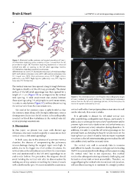
Figure 3. Illustrated cardiac anatomy and surgical procedure of Case 1. B
(A) Illustration depicting cardiac anatomy of Case 1, viewed from the left
posterolateral angle. (B) Illustration depicting anastomosis of dissected
vertical vein with an opening on the left atrial appendage incision.
(C) Illustration depicting completed anastomosis.
Abbreviations: Ao: Aorta; IV: Innominate vein; LAA: Left atrial appendage;
LIPV: Left inferior pulmonary vein; LSPV: Left superior pulmonary vein;
LV: Lingual vein; MPA: Main pulmonary artery; RIPV: Right inferior
pulmonary vein; RSPV: Right superior pulmonary vein; SVC: Superior
vena cava; VV: Vertical vein.
The vertical vein was then opened along its length between
the ligature distally and the silk loop proximally. The dorsal
surface of the left atrial appendage was then opened for a
length of >1 cm (Figure 5B) to correspond to the vertical
vein opening. A wide anastomosis was created between Figure 4. Two-dimensional and color Doppler echocardiography images
these openings using 7-0 polypropylene continuous suture, of Case 1 patient at 6 months follow-up. (A) Unobstructed pulmonary
venous flow into the left atrial appendage (arrow). (B) No flow across the
in a side-to-side fashion (Figure 5C), without disconnecting interatrial septum (indicated by arrow).
the vertical vein from the innominate vein.
The roof of the coronary sinus is split/divided so that vertical wall with a finer polypropylene suture since its wall
the coronary sinus along with the right pulmonary venous can be relatively thin even in adults.
drainage now drains into the left atrium. Echocardiography It is advisable to dissect the left-sided vertical vein
revealed no blood flow turbulence in the vertical vein-left after establishing cardiopulmonary bypass, particularly in
atrial appendage anastomosis. adults, due to severe pulmonary artery hypertension and to
prevent right ventricular dysfunction and arrhythmias as a
3. Discussion result of pulmonary artery retraction during dissection. In
In this paper, we present two cases with distinct age addition, it is safer to open the left atrial appendage on the
difference, who were treated surgically to anastomose their arrested heart, as clamping its base for anastomosis on the
vertical veins to left atrial appendage. beating heart can distort it and is likely to injure important
In both cases, due to the absence of a common venous structures in the left atrioventricular groove such as the left
chamber, the chances of compromising the pulmonary circumflex artery and the coronary sinus.
venous drainage during the surgical repair were high. In The vertical vein wall is extremely thin in neonates
adults, due to the bigger size of all cardiac structures, the and difficult to handle. Recommended approach to treating
vertical vein can be safely disconnected from the innominate PAPVC in neonates involves dividing and then anastomosing
vein and anastomosed to the left atrial appendage in an the vertical vein, but in our experience, this method
5
end-to-side fashion. For precaution, the surgeons should renders the subsequent suturing extremely challenging, and
avoid twisting the vertical vein after its disconnection by formation of any kink or twist unavoidable. Therefore, we
making use of stay sutures or marking the trimmed vessels suggest ligating the vertical vein-innominate vein junction,
with a sterile marker pen. It is recommended to anastomose without disconnecting it, to maintain the straight position
Volume 2 Issue 2 (2024) 3 doi: 10.36922/bh.2376