
Page 95 - BH-2-2
P. 95
Brain & Heart Lipomatous hypertrophy of the septum secundum associated with PFO
A B A B
C D
C D
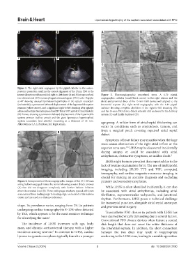
Figure 1. The right atrial angiogram by the pigtail catheter in the antero-
posterior projection confirms the correct alignment of the 15mm FSO to the
interatrial septum with no residual right-to-left shunt. (A and B) Intraprocedural Figure 3. Fluoroangiographic procedural steps. A 6-Fr pigtail
two-dimensional (2D) transesophageal echocardiogram (TEE) color Doppler angiographic catheter (small black arrow) in the right atrium and the
at 44° showing unusual lipomatous hypertrophy of the septum secundum distal and proximal discs of the 15-mm FSO device well aligned to the
(red asterisk), a permanent leftward displacement of the hypermobile septum interatrial septum (A); right atrial angiography with the 6-Fr pigtail
primum (yellow arrow), and a significant right-to-left shunting after agitated catheter showing complete abolition of the right-to-left shunting (B);
saline contrast injection; intraprocedural 2D TEE at 105°, systolic (C) and diastolic and the 15-mm FSO device (black asterisk) still anchored to the delivery
(D) frames, showing a permanent leftward displacement of the hypermobile system (C) and finally deployed (D).
septum primum (yellow arrow) and the giant lipomatous hypertrophied
septum secundum (red asterisk) measuring at a thickness of 25 mm. age group. A milder form of atrial septal thickening can
Abbreviations: LA: Left atrium; RA: Right atrium.
occur in conditions such as amyloidosis, tumors, and
from a surgical patch covering repaired atrial septal
A B
defect.
Symptoms of heart failure may manifest when the large
mass causes obstruction of the right atrial inflow or the
superior vena cava. LHSS may be discovered incidentally
7,8
during autopsy or could be associated with atrial
arrhythmias, obstructive symptoms, or sudden death. 9
LHSS might be more prevalent than reported due to the
lack of routine examination for it. The use of multimodal
imaging, including 2D/3D TTE and TEE, computed
tomography, and cardiac magnetic resonance imaging, is
crucial for making an accurate diagnosis and excluding
Figure 2. Intraprocedural fluoroangiographic images of the 25 × 45 mm primary and secondary neoplasms.
sizing balloon engaged inside the tunnel showing a waist (black arrows)
(A) that did not disappear completely with further balloon inflation While LHSS is often identified incidentally, it can also
above its nominal level (B). Three radiopaque markers, spaced at 10 mm be associated with atrial arrhythmias, including atrial
as measured from leading edge to leading edge, are located at the balloon fibrillation, supraventricular tachycardia, and junctional
center and are used as a distance reference. rhythm. Furthermore, LHSS poses a technical challenge
for transseptal puncture, alongside atrial septal aneurysm
shape. Its prevalence varies, ranging from 2% (in patients and previous atrial surgery.
undergoing cardiac tomography) to 8–10% when detected
by TEE, which appears to be the most sensitive technique Transcatheter PFO closure in patients with LHSS has
for identifying the mass. 5 been deemed technically demanding due to several factors.
Conventional PFO closure devices often feature a central
The incidence of LHSS increases with age, body disk length that does not cover the entire thickness of
mass, and chronic corticosteroid therapy, with a higher the interatrial septum. In addition, the short connection
incidence among women. In contrast to LHSS, cardiac between the two discs may result in inappropriate
6
lipoma is a genuine neoplasm typically found in a younger anchoring to the LHSS rims, leading to unstable apposition
Volume 2 Issue 2 (2024) 3 doi: 10.36922/bh.2190