
Page 100 - BH-2-2
P. 100
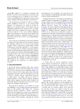
Brain & Heart                                                    Surviving rhino-orbital-cerebral mucormycosis


            uncontrolled diabetes is commonly associated with   documentation was unavailable. On examination, the
            ROCM, a condition that has become more prevalent due   patient exhibited a dilated and fixed left pupil, conjunctival
            to the increasing incidence of diabetes in our society .   chemosis, and periorbital edema.
                                                        2-4
            Diverse vulnerabilities in host immunity result in varying   Imaging tests revealed a left temporal hypodensity on
            degrees of organ involvement and clinical presentations .  contrast computed tomography (CT) (Figure  1C), while
                                                        5-7
              The manifestation of ROCM in COVID-19  patients   contrast magnetic resonance imaging (MRI) demonstrated
            without traditional risk factors is now evident. Early   smooth, regular wall rim enhancement (Figure 1D) with
            diagnosis  and  intervention  are  crucial  to  combat  the   diffusion restriction (Figure  1E), indicating abscess
            aggressive and fulminant nature of this disease and the   formation. The abscess extended into the sphenoethmoidal
            overlapping symptoms with bacterial facial or orbital   air sinuses, gangliocapsular area, and retroorbital region
            cellulitis, which pose a diagnostic challenge . A high index   (Figure  1F), leading to the final diagnosis of stage IV
                                              8
            of clinical suspicion is vital, and a definitive diagnosis   ROCM). An ear swab smear confirmed the presence of
            relies on the presence of non-septate broad hyphae with   aseptate hyphae with right-angle branching, identifying the
            right-angle branching in potassium hydroxide mounts,   pathogen as mucor. Subsequently, the patient underwent left
            lactophenol cotton blue mounts, and histopathological   orbital modified lid-sparing exenteration, resulting in the
            examinations .                                     formation of a fistula (Figure 1G). Despite this intervention,
                       8
              The primary aim of reporting this case was to illustrate   the patient’s condition continued to deteriorate, leading to
            the significant burden experienced by the patient, including   agitation and altered mental status. Consequently, a left
            the challenges endured by the patient during multiple   temporo-parietal craniotomy with abscess excision and
            surgeries, serial complications, psychological distress   augmentation duroplasty was performed. Post-operative
            resulting from living with a ghastly face, mental anguish   contrast CT revealed satisfactory results (Figure 1H). The
            due to social repulsion, the torment of separation from his   biopsy result was suggestive of mucormycosis. Following
            spouse, and the financial and social strain on his caretaker.   surgery, the patient underwent hemimaxillectomy of the
            The secondary aims included reflecting the aggressive and   left maxilla due to disease progression. Despite medical
            challenging nature of the disease, which makes it typically   interventions, the patient was discharged with a disfigured
            troublesome to  recognize  and extremely challenging  to   face (Figure 2A), significantly affecting his personal and
            treat, evaluating the radicality of surgical debridement,   social life. In addition, he experienced a single episode of
            highlighting  the role of  staged reconstruction surgeries,   generalized tonic-clonic seizures, prompting the escalation
            and signifying the importance of multidisciplinary   of antiepileptic medication.
            teamwork.                                            6  months later, the patient experienced another
                                                               seizure episode, necessitating the addition of a second
            2. Case presentation                               antiepileptic drug. However, he subsequently presented
            A 38-year-old patient presented with pain, redness,   with a bogie swelling over the left temporal region, along
            swelling, ptosis, and vision loss in his left eye over the past   with intermittent pus discharge from the previous surgical
            20 days before seeking medical consultation (Figure 1A).   site (Figure 2B). Imaging revealed bone flap osteomyelitis
            He had a history of left upper molar tooth extraction   and an epidural pus collection, necessitating neurosurgical
            5 days before symptom onset and had experienced a past   intervention and a tracheostomy due to microaspirations
            COVID-19 infection without corticosteroid treatment   from palatal perforation (Figure 2C). Sutures were removed
            2  months earlier. The patient reported no history of   10-day post-surgery, and the patient was discharged
            diabetes mellitus or other chronic medical conditions.   (Figure 2D).
            Initial clinical suspicion pointed toward maxillary and   Subsequently, the reconstruction phase began to
            sphenoethmoidal sinusitis with involvement of the medial   improve the cosmetic appearance of the patient’s face.
            orbital wall, leading to orbital cellulitis.       The left orbitonasolabial fistula repair and debridement
              Broad-spectrum antibiotics were initiated, but 4-day   were performed by the plastic surgery team a couple
            post-admission, a necrotic black ulcerative patch appeared   of months after the previous surgery. While planning
            along the left nasolabial fold (Figure  1B), indicating   for reconstructive flap cover surgery during the same
            zygomycete-related  periarteritis,  vascular  thrombosis,   admission, the patient developed a maggot infection at the
            infarction, and angioinvasion. Intravenous amphotericin   apex of the orbit (Figure 2E). Contrast MRI of the brain,
            therapy was promptly initiated. Before presentation at our   orbit, and paranasal sinuses revealed mucosal thickening
            facility, the patient had undergone functional endoscopic   of the left sphenoethmoid and frontal air sinus with
            sinus surgery (FESS) elsewhere, although the relevant   cavernous sinus infiltration and carotid artery encasement


            Volume 2 Issue 2 (2024)                         2                                doi: 10.36922/bh.2083