
Page 101 - BH-2-2
P. 101
Brain & Heart Surviving rhino-orbital-cerebral mucormycosis
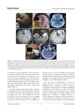
A B C
D E F
G H
Figure 1. Clinical manifestations and surgical outcomes in the case of rhino-orbital-cerebral mucormycosis. (A) Gross periorbital edema, ptosis, and
maxillary fullness on the left side of the face. (B) A black necrotic ulcerative eschar along the left nasolabial fold by the side of the ala of the nose, indicating
fungal etiology (a hallmark of Mucorales infection). (C) Contrast computed tomography (CT) head revealing left temporal hypodensity with well-defined
margins and surrounding edema. (D and E) Axial magnetic resonance imaging (MRI) cuts revealing abscess wall enhancement and diffusion restriction.
(F) Axial contrast MRI cuts revealing extension into left spenoethmoid and retroorbital regions. (G) Post-exenteration figure of the patient with dressing
in situ and formation of fistula due to shredding off of necrotic eschar. (H) Post-operative contrast CT showing complete excision of the abscess cavity with
no residual contrast enhancement.
by necrotic tissue. This complication not only delayed the adjacent to the ala of the nose (Figure 2H). Finally, after
reconstruction surgery but also necessitated debridement 9 months, the day of reconstruction arrived. Debridement
of necrotic tissue and maggot removal (Figure 2F) over of the left orbital necrotic tissue, along with free anterolateral
the left orbital region, along with redo tracheostomy. After thigh flap cover and split skin graft cover (taken from the
surgery, the patient was left with a large cavity, which was left thigh), was performed (Figures 3A and B).
visually displeasing (Figure 2G). He was discharged 4 days Over the course of approximately 26 months, the
later with instructions for a high-protein diet via Ryle’s patient underwent nine major surgical procedures,
tube feeding, tracheostomy care, aseptic dressing, and including extensive reconstructive surgeries. His journey
regular follow-up. was marked with physical and psychological challenges,
The patient needed an additional 9 months to strengthen complications, and significant cosmetic deformities.
his immunity, improve his nutritional status, and clear His older brother played a pivotal role in his care and
all necrotic tissue and residual infestations. However, support, accompanying him through multiple medical
this progress came with a cost. The patient was left with departments. In addition to surgical treatments, he
an asymmetric face, an exenterated left socket, a sagging received liposomal amphotericin and posaconazole to
and sunken scalp flap, and a disagreeable, unsightly, and combat mucormycosis. However, his struggles persist as
gruesome colossal tissue defect over the maxillary area he continues to undergo regular follow-ups in the plastic
Volume 2 Issue 2 (2024) 3 doi: 10.36922/bh.2083