
Page 102 - BH-2-2
P. 102
Brain & Heart Surviving rhino-orbital-cerebral mucormycosis
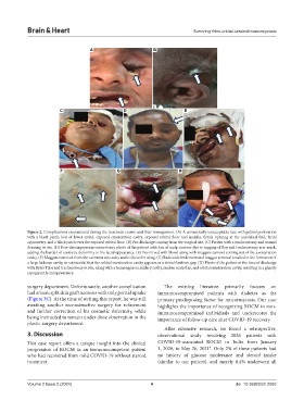
A B
C D E
F G H
Figure 2. Complications encountered during the treatment course and their management. (A) A cosmetically unacceptable face with palatal perforation
with a black patch, loss of lower eyelid, exposed exenteration cavity, exposed orbital floor and maxilla, fistula opening at the nasolabial fold, facial
asymmetry, and a black patch over the exposed orbital floor. (B) Pus discharge coming from the surgical site. (C) Patient with a tracheostomy and wound
dressing in situ. (D) Post-decompression craniectomy photo of the patient with loss of scalp contour due to sagging of flap and tracheostomy scar mark,
adding the burden of cosmetic deformity to his facial appearance. (E) Pus mixed with blood along with maggots (arrow) coming out of the exenteration
cavity. (F) Maggots removed from the exenteration cavity and collected in a bag. (G) Radical debridement and maggot removal resulted in the formation of
a large hideous cavity, so substantial that the orbital exenteration cavity appears as a trivial fusiform gap. (H) Photo of the patient at the time of discharge
with Ryle’s Tube and tracheostomy in situ, along with a humongous maxillary cavity, sunken scalp flap, and orbital exenteration cavity, resulting in a ghastly
repugnant facial appearance.
surgery department. Unfortunately, another complication The existing literature primarily focuses on
had arisen: split skin graft necrosis with only partial uptake immunocompromised patients with diabetes as the
(Figure 3C). At the time of writing this report, he was still primary predisposing factor for mucormycosis. Our case
awaiting another reconstructive surgery for refinement highlights the importance of recognizing ROCM in non-
and further correction of his cosmetic deformity, while immunocompromised individuals and underscores the
being instructed to remain under close observation in the importance of follow-up care after COVID-19 recovery.
plastic surgery department.
After extensive research, we found a retrospective,
3. Discussion observational study involving 2826 patients with
This case report offers a unique insight into the clinical COVID-19-associated ROCM in India from January
9
progression of ROCM in an immunocompetent patient 1, 2020, to May 26, 2021 . Only 2% of these patients had
who had recovered from mild COVID-19 without steroid no history of glucose intolerance and steroid intake
treatment. (similar to our patient), and merely 0.4% underwent all
Volume 2 Issue 2 (2024) 4 doi: 10.36922/bh.2083