
Page 66 - GPD-1-1
P. 66
Gene & Protein in Disease A novel USH2A gene mutation in retinitis pigmentosa
(USH2A) gene . Therefore, this study was conducted abnormalities noted in the vestibular function examination.
[4]
to screen and analyze the USH2A gene mutations in a Therefore, the proband was clinically diagnosed as non-
Chinese family with RP. syndromic RP.
2. Case presentation Next, the proband’s mother had light perception in
both her right and left eyes, with yellowish-white opacity
2.1. Clinical findings of the lens. Furthermore, fundus photography revealed
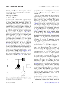
The family in this study has family members who had yellowish waxy optic disks, thin retinal blood vessels,
developed RP. Including the proband, all family members proliferation of a large amount of osteocyte-like pigments
are of Han Chinese ethnic group, and the age of the in the central and peripheral retina, and macular atrophy
proband and her father, mother, and daughter were 55, (Figure 2B). OCT showed the absence of a normal fovea
89, 84, and 29 years, respectively. The proband, who had in the macula of both eyes, with the presence of edema
an approximately 37-year history of night blindness, was between the layers and hyper-reflective continuum of the
diagnosed with non-syndromic RP, whereas her mother epiretinal membrane. Further, both the outer nuclear layer
was diagnosed with type II Usher syndrome. In contrast, and the nerve fibers around the optic disk were thinned
her father and daughter had no clinical manifestations of (Figure 2D and F). In contrast to the proband, her mother
RP and presented with a normal phenotype (Figure 1). The showed sensorineural high- and low-frequency hearing
proband had a history of night blindness of 18 years with loss in both ears, as shown in Figure 2H according to
no symptoms of hearing loss. She had a visual acuity level the pure-tone audiometry examination. However, no
of 0.1 and 0.2 in the right and left eyes, respectively. Fundus obvious abnormality was noted in the vestibular function
photography revealed a yellowish waxy optic disk, thin examination. Thus, the proband’s mother was clinically
retinal blood vessels, and proliferation of a large amount diagnosed as type II Usher syndrome. In contrast, no
of osteocyte-like pigments in the central and peripheral abnormality was noted in the ophthalmic, vestibular
retina. In addition, atrophic changes and proliferative function, and pure-tone audiometry examination in the
membranes were observed in the macula, as shown in proband’s father and daughter.
Figure 2A. Optical coherence tomography (OCT) showed 2.2. Identification of the USH2A gene mutations
the absence of a normal fovea in the macula of both eyes,
with the presence of cystoid edema between the layers and Whole-exome sequencing of the genomic DNA of the
hyper-reflective continuum of the epiretinal membrane. proband led to the identification of two mutations in the
Further, both the outer nuclear layer and the nerve fibers USH2A gene, namely, the heterozygous variants c.8559-
around the optic disk were thinned (Figure 2C and E). 2A>G and c.151A>T (p.Ile51Phe) (NM_20 6933), which
Pure-tone audiometry subsequently confirmed that her were compared with the UCSC hg19 human genome
high- and low-frequency hearing abilities were within the reference sequence to identify the genetic variations.
normal range, as shown in Figure 2G, with no obvious On average, the mean coverage of the target regions was
182.13X, and for each sample, more than 99.77% of target
regions were covered. Sanger sequencing for the identified
mutations was performed, and the presence of these two
variations in the proband family members was, further,
determined and verified. The verification results were
consistent with the Illumina sequencing results, as shown
in Figure 3. The c.8559-2A>G genetic mutation has been
reported previously in multiple studies. Interestingly
c.151A>T (p.Ile51Phe) is a novel genetic mutation which
was identified for the first time. The c.151A>T (p.Ile51Phe)
variant located at chr1-216595528 with a nucleotide
switch, that is, A to T at position 151 of the second exon
of the USH2A gene, results in an amino acid change
from isoleucine to phenylalaninat at position 51 of the
corresponding peptide.
2.3. Pathogenicity analysis of the gene mutations
Figure 1. Retinitis pigmentosa pedigree. ■: Male patient; ●: Female
patient; □: Healthy male; ○: Healthy female; ↗: Proband; : Retinitis The c.8559-2A>G variant, known as splice-site mutation,
pigmentosa (RP) combined with hearing loss. is located at chr1-216051224. It has an allele frequency of
Volume 1 Issue 1 (2022) 2 https://doi.org/10.36922/gpd.v1i1.106