
Page 67 - GPD-1-1
P. 67
Gene & Protein in Disease A novel USH2A gene mutation in retinitis pigmentosa
A B
C D
1 1
C D
2 2
E 1 F 1
E F
2 2
G H
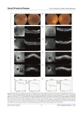
Figure 2. Clinical data of the proband (left) and her mother (right). Fundus photography (A and B), optical coherence tomography (OCT) of the macula
(C and D), thickness of the nerve fibers around the optic disk (E and F), and pure-tone audiometry (G and H). The pictures (C , D , E , F ) represent the
1
1
1
1
right eyes, and pictures (C , D , E , F ) the left eyes. Fundus photography showed that the optic disk was yellowish and waxy, the retinal blood vessels were
2
2
2
2
thin, and a large number of osteocyte-like pigments proliferated in the central and peripheral retina. OCT of the macula revealed the absence of a normal
fovea in the macula of both eyes, with cystoid edema between the layers. Hyper-reflective continuum of the epiretinal membrane was visible. Both the
outer nuclear layer and the nerve fibers around the optic disk were thinned. Pure-tone audiometry indicated that the hearing of both ears of the proband
was within the normal range. In contrast, her mother had hearing loss at both the high- and low-frequencies in both the ears.
Volume 1 Issue 1 (2022) 3 https://doi.org/10.36922/gpd.v1i1.106