
Page 72 - GPD-3-2
P. 72
Gene & Protein in Disease Opportunities and challenges of HIF-1 in cancer
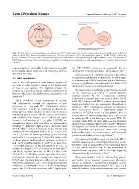
Figure 2. Under hypoxic conditions, hypoxia-inducible factor-1 (HIF-1) translocates to the nucleus, where it targets genes encoding vascular endothelial
growth factor (VEGF), transforming growth factor-alpha (TGF-α), erythropoietin (EPO), phosphoglycerate kinase 1 (PGK1), and BCL2 interacting
protein 3 (BNIP3). These genes affect erythropoiesis, angiogenesis, and metabolism through the hypoxia response element (HRE). The expression of
BNIP3 induces mitophagy, where mitochondria are engulfed by autophagosomes, mitigating stress and maintaining energy production despite hypoxic
conditions.
response highlights the need for further research, especially the HIF-1/STAT3 interaction is responsible for the
in mitigating cancer resistance and developing effective development of immunoresistance in lung cancer cells. 45
anticancer therapies. Toll-like receptor 4 (TLR4) is a member of the pattern
2.2. HIF-1 interactions recognition receptor family of proteins and an HIF-1 target.
Its interaction with HIF-1 contributes to the inflammatory
Due to the rapid growth of solid tumors, oxygen and process in glioblastoma tumorigenesis and leads to the
46
nutrients are often depleted, leading to the development development of pancreatic adenocarcinoma. 47
of hypoxia and necrosis. This depletion triggers the
production of proinflammatory mediators, recruitment of The preservation of the Warburg effect heavily depends
immune cells, tumor cell proliferation, angiogenesis, and on the expression and activity of several glycolytic
metastasis. 38 enzymes activated by HIF-1. Hexokinases, aldolases,
6-phosphofructokinase liver type, enolase alpha, PKM2,
HIF-1 contributes to the maintenance of hypoxia and LDH-A interact with HIF-1 to ensure uninterrupted
and inflammation through the regulation of gene energy production and macromolecular biosynthesis in
expression of c-Jun and AP-1 transcription factors. malignant cells. Collaboration between the oncogenic
48
This regulation provides an additional mechanism for MYC transcription factor, a mammalian target of
tumor growth, which involves the induction of vascular rapamycin, and HIF-1 results in a high glycolytic flux. As
endothelial growth factor (VEGF), tyrosine hydroxylase, a consequence of enhanced glycolysis, there is an increase
and endothelin-1 in hypoxic areas. NF-κB and signal in lactate and H levels, leading to an acidic TME. The
39
+
transducer and activator of transcription 3 (STAT3) are resulting acidic TME, in turn, attenuates the antitumor
transcription factors that contribute to inflammatory immunity and reduces drug uptake by tumors. Moreover,
49
signaling. 40,41 Inhibitory IκB proteins dissociate from the hypoxic conditions in pancreatic cancer stimulate
NF-κB, which, further, translocates to the nucleus and HIF-1-dependent tumor–stromal interactions, creating
activates pro-survival genes such as BCL2, CXCR1, and a fibroinflammatory microenvironment with decreased
CXCR2, as well as tumor-promoting genes encoding perfusion and limited cancer drug delivery. 50
interleukin-6, cyclooxygenase 2, inducible nitric oxide
synthase, platelet endothelial cell adhesion molecule-1, One study highlighted the important role of the HIF-1/
and matrix metalloproteinase-9 (MMP-9), as part of an carbonic anhydrase IX system in the production of soluble
orchestrated response to the inflammatory process, as mediators, such as granulocyte colony-stimulating factor
illustrated in Figure 3. 40,42 The interaction between HIF-1 (G-CSF), which are necessary for the recruitment of
and STAT3 regulates the activity of MCL1, c-Myc, cyclin myeloid-derived suppressor cells to the lungs, triggering
51
D1, MMP-2, and VEGF, which are responsible for survival, the generation of premetastatic niches.
proliferation, invasion, and angiogenesis, respectively. The process in which epithelial cells lose their polarity
43
STAT3 also physically interacts with HIF-1α, which is and switch to an invasive mesenchymal phenotype,
vital for the activation of HIF-1 target genes under hypoxic known as epithelial-to-mesenchymal transition (EMT),
conditions, as illustrated in MDA-MB-231 human breast significantly impacts tumor metastasis. HIF-1 and TGF-β
52
cancer and RCC4 renal carcinoma cells. Significantly, mutually enhance each other’s expression and provoke
44
Volume 3 Issue 2 (2024) 5 doi: 10.36922/gpd.3431