
Page 94 - GPD-4-1
P. 94
Gene & Protein in Disease Oral-ERT in PD knockout mice with tobrhGAA
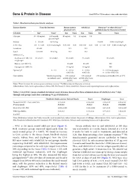
Table 1. Biochemical/enzyme kinetic analyses
Enzyme kinetics From the literature Human mature tobrhGAA Myozyme** or other rhGAAs**
placental GAA published data for Myozyme is limited
Substrate Km* Vmax* Km Vmax Km Vmax Km Vmax
Glycogen 10 – 42 mg/mL 6.63 mmole 18 mg/mL 5.2 21 mg/mL 5.8
glucose/mg/h
Maltose 2 – 22 mM 14.3 U/mg 16 mM 0.186 mmol/mg/h
4-MU-Glyc 0.8 – 1.1 mM 0.55 mmole/mg/h 0.95 mM 0.85 0.85 mM 0.65 1.08 – 2.1 mM 0.07 – 0.548 mmole/mg/h
Isomaltose 40 mM ND ND
Starch 15.4 mM 10 U/mg ND ND
Inhibitors IC 50
Acarbose on 4-MU-Glc 81 umol/L 54 umole/L 85 umol/L 75 umol/L 59 umol/L
on glycogen
Maltose on 4-MU-Glc 45 mM 45 mM ND
Glycogen on 4-MU-Glc 15 mg/mL 10 mg/mL ND
Optimal pH 4 – 5.5 4 – 4.5 for 4 – 4.5 for 4 – 4.25 for glycogen
4-MU-Glc 4-MU-Glc 4 – 5.5 for 4-MU-Glc
Heat stability Variable depending <5% residual <5% residual <5% residual activity after 24 h at 37°C
on methods used activity after 5 min activity after 5 min
at 56°C at 56°C
Notes: *From literature for various species and tissue sources; **Published data for Myozyme is limited.
Abbreviations: GAA: Acid a-glucosidase; h: Hour; ND: Not found; U: Units; tobrhGAA: Tobacco seeds expressing human acid a-glucosidase.
Table 2. GAA/NAG (mean±standard deviation) assay of mouse tissues after three administrations of tobrhGAA over 7 days
through oral gavage (each dose containing 75 µg of tobrhGAA)
Hindlimb tibialis anterior Skeletal Muscle Heart Diaphragm Liver
Treated GAA KO - Oral tobrhGAA 0.12±0.04 0.10±0.05 0.13±0.07 0.19±0.02
P versus mock P≤0.015 P≤0.15 P≤0.16 P≤0.0001
Mock GAA KO- PBS 0.043±0.005 0.06±0.008 0.07±0.03 0.05±0.007
Wild-type-Balb/c 1.50±0.2 0.43±0.16 0.77±0.12 1.00±0.11
% of WT 8 23 17 19
Notes: Bold values indicate the P value versus the mock treated; Italic values indicate the percent of wildtype. Abbreviations: GAA: Acid a-glucosidase;
PBS: Phosphate buffered saline; KO: Knockout; WT: Wild-type; tobrhGAA: Tobacco seeds expressing human acid a-glucosidase.
– 95%) to the mock-treated wild-type mice (Figure 4). Serum antibody titer to oral tobrhGAA at 203 days
Both treatment groups improved significantly from the was undetectable at 4 weeks, barely detected at a 1:10 at
mock-treated group (P ≤ 0.007). We found an increase 8 weeks for both 1× and 3× treatments, and detected at
in GAA activity in the tissues (heart, hindlimb skeletal 1:20, thus demonstrating a low immune response. We
100
muscle, kidney, liver, and diaphragm) from the GAA simultaneously generated mouse antibodies to human
KO mice treated with tobrhGAA by oral gavage, thereby placental GAA by subcutaneous injection of 15 µg over
supporting Oral-ERT with tobrhGAA. The improvement 3 months and found the titer to be 1:1500 (data not shown).
percentage compared to the wild-type ranged from 13% to Thus, oral tobrhGAA of a similar antigen quantity did not
38% depending on the tissue (Table 3) (mean ± SD, with generate a high serum immunoreaction. We determined
statistical significance indicated), with the diaphragm whether tobrhGAA was taken up by taking a tissue sample
being the highest at 38%. We also measured the tissue from the tail end without sacrificing the mice and assayed
glycogen levels after 203 days of treatment and found it for GAA at days 45 and 203 (Figure 5). Wild-type mice
109
ranged from 18% to 48% reduction from the mock-treated had GAA activity of 0.46 ± 0.14, mock-treated GAA KO
mice, with the greatest reduction in the skeletal muscle had 0.064 ± 0.021, and treated GAA KO had 0.10 – 0.2
and diaphragm (43% and 48%, respectively) (Table 4) ± 0.05 for the 1× or 3× treatments (P ≤ 0.006 treated vs.
(mean ± SD, with statistical significance indicated). mock-treated).
Volume 4 Issue 1 (2025) 8 doi: 10.36922/gpd.1760