
Page 20 - GTM-3-1
P. 20
Global Translational Medicine Role of HTS in cancer therapeutics
(2) fluorescence polarization assay). Therefore, labeling of compounds without altering their phenotypic
166
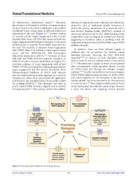
identification of biomarkers of these reprogramming at properties, that is, multipolar spindle formation to
the level of gene and protein could help to explain MOA profile the cellular interactome of a molecule with a
of different tumor variant types in different individuals non‑covalent binding profile. PARPYnD retained its
depending on the case (Figure 3). Another method phenotypic behavior and in vitro PARP binding profile
169
to measure cellular target engagement is the Cellular comparable to parent compounds AZ9482 and AZ0108,
Thermal Shift Assay (CETSA) that measures label‑free suggesting its beneficial utility in profiling novel off-
target engagement with endogenous protein. In this assay, target interactions of both AZ0108 and the clinical PARP
antibody pairs to quantify thermostable target proteins inhibitor olaparib.
are used. For example, to measure target engagement
with PARP1, Shaw et al. utilized a triple-negative breast In addition, there are other relevant targets or
cancer cell line, MDA‑MB‑436, with homozygous pathways that can be assessed, for instance, cancer
deleterious mutations in BRCA1. Treatment of these signaling pathways involving the p53, RTK–RAS
170
cells with the PARP inhibitors olaparib, rucaparib, or signaling 172-174 or various cell-cycle regulation targets
NMS‑P118 led to a thermal stabilization of roughly 2°C, such as cyclin D, cyclin E, and the mitotic kinase Aurora
providing evidence of target engagement with cellular 2. 175,176 Mortality rate is higher in breast cancer patients
PARP1. CETSA can be used for bridging the gap between who overexpressed cyclin-dependent kinase 2/cyclin
target engagement and the desired functional effect. E2 (CDK2/cyclin E2) through the estrogen receptor
Pharmacological inhibition of PARP1 with olaparib pathway. 177,179 Another serine/threonine kinase oncogene,
was also determined by another approach in a study by STK15/BTAK (approved gene symbols are AUR2, ARK1,
Howard et al., where they demonstrated the application and AIK1) required for the formation of the mitotic
of PARPYnD, the first photoaffinity‑based probe (AfBP) bipolar spindle, has been reported to be overexpressed
177
for profiling PARP1/2. This approach was used to in breast cancers. These targets could be used in future
171
profile clinical PARP inhibitor olaparib and to identify to find more potent and selective cancer drugs. However,
off-target proteins. This strategy utilizes photoaffinity a study has shown that targeting Aurora proteins
171
Figure 3. Promising biological targets for cancer drug screening.
Volume 3 Issue 1 (2024) 12 https://doi.org/10.36922/gtm.2448