
Page 105 - GTM-3-4
P. 105
Global Translational Medicine Prediction of in-stent restenosis
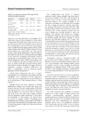
Table 3. Cox regression evaluation of the impact of risk The vascular-related risk factors of coronary
factors for coronary restenosis restenosis include complex calcified, long vessel lesions,
atherosclerotic plaques in the Ostia, and bifurcations of
Risk factor Coefficient HR 95% CI P coronary arteries, as well as small vessel diameter and
Male sex 0.785 2.194 1.5 – 3.22 <0.001*** multivessel disease. For example, Coughlan et al.
29
30
Prior myocardial 0.0934 1.098 1.05 – 1.15 0.045* presented a novel clinical score (the ISAR score) to predict
infarction the risk of repeat percutaneous coronary intervention
Nominal stent −0.338 0.713 0.58 – 0.87 0.0011** for recurrent DES-in-stent restenosis (DES-ISR). They
diameter (<2.5 mm) retrospectively analyzed 1,986 consecutive patients with
Stent type (BMS) −0.591 0.554 0.41 – 0.75 0.0001*** DES-ISR (2,392 in-stent restenosis lesions) from two
Note: *P<0.05; **P<0.01; ***P<0.001. centers. Patients were randomly divided (3:1 ratio) into
Abbreviations: BMS: Bare metal stent; HR: Hazard ratio; training (1,471 patients, 1,778 lesions) and validation
CI: Confidence interval.
(515 patients, 614 lesions) cohorts to develop and validate
the predictive model. The median duration of clinical
restenosis is also associated with a much higher rate of follow-up after DES-ISR treatment was 7.4 years. Four
failure than revascularization treatment of native vessels. clinical variables were associated with repeat percutaneous
Consequently, DES significantly reduces the incidence of coronary intervention for recurrent DES-ISR after 1 year:
in-stent restenosis; however, the problem remains relevant. (i) A non-focal in-stent restenosis pattern, (ii) time interval
The currently known risk factors for in-stent restenosis to in-stent restenosis <6 months, (iii) in-stent restenosis
can be classified into three groups: patient-related, vessel- in the left circumflex coronary artery, and (iv) in-stent
related, and procedure-related. Patient-related risk factors restenosis in a calcified vessel. In addition, the numerical
18
include patient age, comorbidities (e.g., DM), genetic four-item ISAR score (one point for each variable) proved
disorders, and systemic inflammation, with DM being the clinically useful to readily predict percutaneous coronary
most important risk factor in this group that may trigger intervention for recurrent DES-ISR.
in-stent restenosis. According to the Swedish Coronary
Angiography and Angioplasty Registry (SCAAR), which Percutaneous coronary intervention-related risk
includes information about 35,000 stented patients, after factors include stent deployment failure, excessive stent
2 years of follow-up, restenosis in patients with DM was dilatation, stent fracture, and polymer damage. These cases
twice as frequent with the zotarolimus-eluting stent. occur most frequently when stenting complex lesions
19
This is due to metabolic disorders that trigger endothelial or markedly calcified vessels, resulting in neointimal
dysfunction, accelerate neointimal proliferation, and hyperplasia and impaired delivery of the drug coating
31
induce a prothrombotic state by increasing platelet the stent. Some investigators proposed new biomarkers,
aggregation and thrombogenicity. 20,21 such as the triglyceride-glucose index and epicardial fat
32
thickness. 33
Several papers demonstrate the role of genetic
abnormalities, such as polymorphisms of genes encoding Coronary restenosis remains a common complication
haptoglobin 2/2.25, interleukin-8 (IL-8), angiotensin- following coronary revascularization and stenting. The
converting enzyme, and glycoprotein receptor IIIaPLA1/2, technical reasons for in-stent restenosis are well elucidated.
in the development of stent restenosis in most of these Multiple studies indicated that adequate stent deployment,
patients. 22-24 complete inflation, and optimal stent diameter play key
roles in preventing restenosis. In addition, the complexity
Multiple studies have been conducted to identify
inflammation markers and correlate their levels with of the procedure due to multivessel disease, bifurcation
lesions, small-diameter vessel lesions, and pronounced
coronary restenosis. Plasma C-reactive protein (CRP) level
has long been proven to be a CVD predictor and is now calcification is another important risk factor.
being investigated for its predictive value for restenosis. Our study analyzed known predictors of coronary
One study found that CRP levels strongly correlated restenosis in a large sample of stented patients who
with the angiographic signs of restenosis. 25,26 The number developed hemodynamically significant restenosis,
of macrophages in tissue samples and restenosis have requiring repeat revascularization within 60 months of the
been found to strongly correlate. Circulating matrix first stenting. The roles of sex and history of myocardial
27
metalloproteinases (MMPs), namely, MMP-2 and MMP-9, infarction have been demonstrated for restenosis
have recently been identified as potentially useful markers formation and the rate of its development. Moreover,
for identifying patients at high risk of restenosis after stent DES is significantly superior to BMS in terms of adverse
implantation. 28 event incidence and rate. These results are consistent with
Volume 3 Issue 4 (2024) 7 doi: 10.36922/gtm.4957