
Page 12 - GTM-4-3
P. 12
Global Translational Medicine Inflammation in CVD: Mechanisms and markers
In immune regulation, CD47, widely expressed on
vascular endothelial cells, macrophages, and platelets,
plays a crucial role by interacting with signal regulatory
protein-alpha (SIRPα) on phagocytes. This interaction
7
generates a “do not eat me” signal that suppresses
macrophage-mediated clearance of apoptotic cells. In the
context of ASCVD, increased CD47 expression within
plaques promotes immune evasion, allowing damaged
endothelial and lipid-laden foam cells to persist, fueling
chronic inflammation and plaque progression. Impaired
7
efferocytosis leads to necrotic core expansion, further
increasing the risk of plaque rupture and thrombosis. This
dysfunction is mediated through the thrombospondin-1/
CD47/SIRPα signaling, which limits apoptotic
cell clearance and enhances IL-1β release through
inflammasome activation, amplifying vascular immune
responses. Elevated CD47 levels correlate with plaque
7
vulnerability, supporting its potential role in inflammatory
risk stratification. Preclinical studies have demonstrated
that CD47 blockade restores efferocytosis and reduces
plaque burden, underscoring its emerging relevance as a
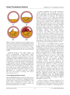
Figure 2. Four stages of atherosclerosis, from fatty deposition to plaque prognostic biomarker and therapeutic target in ASCVD.
rupture: (i) Early fatty streak formation: Initial accumulation of lipids in Besides, SGK1 is a serine/threonine kinase involved
the arterial wall. (ii) Plaque expansion: Increased lipid accumulation with in ion transport, cellular survival, and inflammatory
inflammatory cell infiltration. (iii) Advanced plaque: Plaque enlargement
with cholesterol deposits and immune cell involvement. (iv) Plaque signaling. In ASCVD, it contributes to vascular remodeling
rupture and thrombosis: Ulceration and rupture of the plaque, leading by modulating endothelial function, promoting smooth
to potential clot formation and vessel occlusion. Image was created using muscle proliferation, and activating immune cells. SGK1
8
Notability. enhances NF-κB signaling and NACHT, LRR, and PYD
Additional biomarkers offer further insights into domains-containing protein 3 (NLRP3) inflammasome
ASCVD pathophysiology. Myeloperoxidase (MPO) activation, leading to increased IL-6 and IL-1β production
released by inflammatory cells catalyzes LDL and HDL and subsequent leukocyte recruitment. It also disrupts
oxidative modifications, impairing cholesterol efflux and endothelial nitric oxide synthase activity, promoting
contributing to plaque instability. Lipoprotein-associated endothelial dysfunction and impaired vasodilation.
5
phospholipase A2 (Lp-PLA2), primarily associated with In advanced plaques, SGK1-driven smooth muscle
LDL particles, promotes oxidative stress and the progression proliferation promotes neointimal thickening and
of atherosclerotic plaques by releasing pro-inflammatory arterial stiffening, exacerbating the hemodynamic burden
mediators. In large cohort studies, elevated plasma levels of atherosclerosis. Inhibition of SGK1 in preclinical
of Lp-PLA2 have been independently linked to coronary models has been shown to suppress NF-κB activity,
artery disease events. Finally, trimethylamine-N-oxide, a reduce caspase-1 activation, and dampen downstream
5
gut microbiome-derived metabolite, exacerbates vascular cytokine production, resulting in attenuation of cardiac
8
inflammation by promoting cholesterol deposition inflammation and remodeling. SGK1 expression is also
and foam cell formation, correlating with adverse elevated in thrombi from patients with acute myocardial
cardiovascular outcomes. infarction, suggesting potential utility in identifying
high-risk inflammatory states. These findings highlight
2.5. Emerging biomarkers in ASCVD SGK1 as a mechanistic driver of vascular inflammation
While established biomarkers, such as hs-CRP and IL-6, and a promising biomarker and therapeutic target in
help assess systemic inflammation, emerging biomarkers inflammation-mediated CVD.
provide deeper insight into disease mechanisms. These Similarly, P-selectin, an adhesion molecule stored in
include cluster of differentiation (CD)47, serum and the α-granules of platelets and Weibel–Palade bodies of
glucocorticoid-regulated kinase 1 (SGK1), P-selectin, and endothelial cells, is rapidly translocated to the cell surface
growth differentiation factor 15 (GDF15). on activation, where it binds to P-selectin glycoprotein
Volume 4 Issue 3 (2025) 4 doi: 10.36922/GTM025100023