
Page 226 - IJB-10-1
P. 226
International Journal of Bioprinting 3D printing of costal cartilage models
imported into Mimics for 3D reconstruction, resulting in
the generation of STL files. The newly constructed 3D model
files and the original digital model files were both input into
Geomagic Control 2014 software (Geomagic, NC, USA).
The initial digital model was set as the reference, while the
newly built 3D reconstruction model was set as the test.
The best-fit alignment method was used for automatic 3D
matching, and we analyzed the morphological similarity
of the two models by comparing the 3D deviation using
deviation chromatograms. The same method was used for
intergroup comparison of personalized printed models to
evaluate the precision of the models.
2.6. 3D printing costal models for clinical practice
(ear framework handcrafting curricula and
pre-operative planning)
We assessed resident confidence in 10 learners who tried
3D-printed models using a retrospective scoring system. 51,52
Briefly, the learners rated their pre-training and post-
training confidence levels after seven surgical simulations
on a scale of 1 to 5, reflecting their confidence level changes
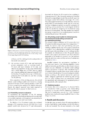
Figure 2. S300 3D printer. Carriers for storing silicone component A (A), and skill gains after practicing on costal cartilage models.
component B (B), and supporting material (C). Printing nozzles of the To evaluate the effectiveness of the 3D-printed silicone
mixed silicone (D) and supporting material (E). Costal cartilage model
(F) and supporting structure (G) in printing. models, each surgeon was asked to score their subjective
impression of the models on a scale of 1 to 5 for hardness,
elasticity, stickiness, suture-ability, and overall satisfaction,
software, and the molding direction and position of
the model were adjusted; as shown in Table 1.
Enrolled patients for pre-operative simulation in
(5) An extrusion nozzle of 0.4 mm and appropriate the study were based on the following eligibility criteria:
process parameters, such as scanning speed of unilateral microtia, good health, no other chronic diseases
15 mm/s and layer thickness of 0.2 mm, were except auricular deformity, and healthy costal cartilage
selected for parametric slicing using FAMufacture, without calcification or trauma. After manufacturing the
with a 100% filling rate and 80% material supply 3D-printed silicone models, three senior plastic surgeons
rate, using the orthogonal scanning strategy [0°, 90°] were invited to handcraft ear frameworks using the models,
for interlayer filling, directly slicing in FAMufacture, mainly according to the method reported in a previous
generating the Gcode print file, and then carrying study. Surgeons optimized the harvesting and carving
53
out 3D printing.
plan for costal cartilage based on personalized models
After completion of the printing process, the printed pre-operatively and implemented the optimized protocol
silicone costal cartilage models were heated in a drying intraoperatively with maximal effort.
oven for 30–60 min at 80°C and then heated to 120°C for
15 min to complete post-curing of the printed constructs. 2.7. Statistical analysis
Finally, the support materials were easily removed by Continuous data were analyzed by the Kruskal‒Wallis
flushing with water to prepare the final construct. test (mechanical tests) or paired t-test (handcrafting
curricula) or one-way ANOVA (Subjective evaluation).
2.5. Printability outcome evaluation The level of significance was set at p < 0.05. Statistical
To demonstrate the printability of the 3D printing analyses were conducted using GraphPad Prism 9.4 (San
technology used in this study, geometries with varying levels Diego, CA, USA).
of structural complexity were examined, encompassing the
production of auricular and nasal models. 3. Results and discussion
The fidelity of the 3D-printed models was evaluated For the first time, we used a direct 3D-printing method to
through a 3D comparison process. DICOM files of each produce high-fidelity costal cartilage models with silicone
printed model were obtained by CT and subsequently as the raw material. The silicone materials in this study
Volume 10 Issue 1 (2024) 218 https://doi.org/10.36922/ijb.1007