
Page 224 - IJB-10-1
P. 224
International Journal of Bioprinting 3D printing of costal cartilage models
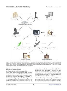
Figure 1. Schematic diagram of the fabrication and application of 3D-printable (3DP) silicone costal cartilage models. The discarded costal cartilage
collected from the operation was matched with the printable silicone material according to the results of mechanical tests. Computed tomography (CT)
scanning and 3D reconstruction provided personalized digital models of costal cartilage. The printed silicone models can be used for pre-operative
simulation, surgical teaching, and receiving subjective evaluation feedback.
2. Material and methods cartilage pieces were then immersed in 0.9% NaCl solution
and stored at -80°C until testing. To ensure complete
48
2.1. Materials and human tissue collection thawing and stress equilibration, the samples were thawed
Native costal cartilage pieces obtained from discarded before testing and then cut into their final shape. Using
postoperative tissue were used as the control material, and discarded postoperative tissue is a sustainable and ethical
cartilage without calcification was included according to approach to using medical waste materials.
pre-operative computed tomography (CT) scanning. To
obtain costal cartilage samples with desirable properties, Three different types of two-component 3DP silicones
the middle portion of the cartilage was utilized, while the and soluble supporting material were used in this study.
perichondrium was removed as thoroughly as possible. The The materials were provided by Elkem Silicones, Shanghai,
Volume 10 Issue 1 (2024) 216 https://doi.org/10.36922/ijb.1007