
Page 219 - IJB-10-1
P. 219
International Journal of Bioprinting CS-laden microporous bio-ink for cartilage regeneration
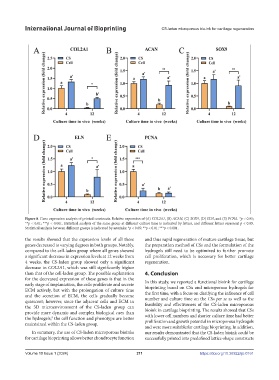
Figure 8. Gene expression analysis of printed constructs. Relative expression of (A) COL2A1, (B) ACAN, (C) SOX9, (D) ELN, and (E) PCNA. *p < 0.05;
**p < 0.01; ***p < 0.001. Statistical analysis of the same group at different culture time is indicated by letters, and different letters represent p < 0.05.
Statistical analysis between different groups is indicated by asterisks: *p < 0.05; **p < 0.01; ***p < 0.001.
the results showed that the expression levels of all these and thus rapid regeneration of mature cartilage tissue, but
genes decreased to varying degrees in both groups. Notably, the preparation method of CSs and the formulation of the
compared to the cell-laden group where all genes showed hydrogels still need to be optimized to further promote
a significant decrease in expression levels at 12 weeks from cell proliferation, which is necessary for better cartilage
4 weeks, the CS-laden group showed only a significant regeneration.
decrease in COL2A1, which was still significantly higher
than that of the cell-laden group. The possible explanation 4. Conclusion
for the decreased expression of these genes is that in the In this study, we reported a functional bioink for cartilage
early stage of implantation, the cells proliferate and secrete bioprinting based on CSs and microporous hydrogels for
ECM actively, but with the prolongation of culture time the first time, with a focus on clarifying the influence of cell
and the secretion of ECM, the cells gradually become number and culture time on the CSs per se as well as the
quiescent; however, since the adjacent cells and ECM in feasibility and effectiveness of the CS-laden microporous
the 3D microenvironment of the CS-laden group can bioink in cartilage bioprinting. The results showed that CSs
provide more dynamic and complex biological cues than with lower cell numbers and shorter culture time had better
the hydrogels, the cell function and phenotype are better proliferation and growth potential in microporous hydrogels
8
maintained within the CS-laden group.
and were more suitable for cartilage bioprinting. In addition,
In summary, the use of CS-laden microporous bioinks our results demonstrated that the CS-laden bioink could be
for cartilage bioprinting allows better chondrocyte function successfully printed into predefined lattice-shape constructs
Volume 10 Issue 1 (2024) 211 https://doi.org/10.36922/ijb.0161