
Page 214 - IJB-10-1
P. 214
International Journal of Bioprinting CS-laden microporous bio-ink for cartilage regeneration
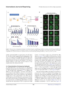
Figure 4. Characterization of microporous hydrogels. (A) Schematic diagrams, optical images, rhodamine B staining, and SEM images of GelMA+PEO
(microporous hydrogel) and GelMA. (B) The pore size analysis. (C) The porosity analysis. (D) Swelling ratio. (E) Viscosity–shear rate curve. (F) Modulus–
temperature curve. (G) Stress–strain curve. (H) Stress–strain curve (0%–30% strain). (I) Young’s modulus. *p < 0.05; **p < 0.01; ***p < 0.001.
and the continued death of a small number of cells within number of micropores (dark areas indicated by white
CS. The above results suggested that the enlargement of the arrows in Figure 4A) in GelMA (red fluorescence, emitted
CSs in non-adherent microwells with culture was due to the by the hydrogel conjugated with rhodamine B). The results
continuous secretion of ECM by chondrocytes rather than of SEM further confirmed that numerous micropores were
cell proliferation and that the cell number of CSs had no formed in the GelMA+PEO group, and it is noteworthy
obvious influence on the GAG secretion function of the that many channels were formed on the walls of these
chondrocytes if the cells survived well. micropores that could connect to the adjacent micropores
(as indicated by yellow arrows in Figure 4A). The pore size
3.3. Characterization of microporous hydrogels and porosity analysis revealed that the GelMA+PEO group
As mentioned above, the dense texture of conventional (72.61 ± 5.30 μm and 63.62% ± 2.25%) had significantly
hydrogels is an important factor hindering the establishment larger pore size and higher porosity than the GelMA group
of cell–cell/cell–ECM interactions. 22,24 To address this (3.50 ± 0.54 μm and 45.80% ± 1.172%; Figure 4B and C).
dilemma, in this study, PEO, a non-toxic inert polymer, Since appropriate swelling, rheological, and mechanical
was introduced into GelMA to prepare the microporous properties are critical for extrusion 3D printing, these
hydrogel based on the phase separation void-formation properties of microporous hydrogel (GelMA+PEO) were
strategy. Due to the immiscibility of the PEO and the evaluated. For swelling ratio, the results showed that the
GelMA solutions, the PEO droplets can be leached off after swelling ratio was 108.1% ± 0.56% for GelMA and 111.6%
the GelMA crosslinking and consequently form numerous ± 2.46% for GelMA+PEO, while there was no statistical
micropores in the cured hydrogels. Rhodamine B staining difference between the two groups (Figure 4D). For the
showed that the introduction of PEO can generate a large rheological properties, it can be seen from Figure 4E
Volume 10 Issue 1 (2024) 206 https://doi.org/10.36922/ijb.0161