
Page 210 - IJB-10-1
P. 210
International Journal of Bioprinting CS-laden microporous bio-ink for cartilage regeneration
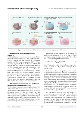
Figure 1. Schematic illustration of the process of non-adhesive microwells preparation and CSs formation.
2.5. Preparation of GelMA with microporous The swelling test was carried out by immersing the
structures cured hydrogel samples in DPBS for 24 h at 37°C and
GelMA with microporous structures was prepared using recording the change in weight of the samples. The swelling
previously established protocols. First, lyophilized GelMA ratio is calculated using the following formula:
26
and PEO powder were fully dissolved in the complete
medium at 60°C to a final concentration of 10% (w/v) and Swelling ratio = W swelling /W × 100% (I)
0
1% (w/v), respectively. Then, the dissolved GelMA+PEO
solution was sterilized by pasteurization and stored at where W swelling is the weight of the hydrogel samples after
-20°C in the dark. Before use, LAP was added to a final swelling in DPBS, and W is the initial weight of the
0
concentration of 0.25%, and blue light was used to induce hydrogel samples.
photocrosslinking of pre-gel solution (wavelength: 405 nm; Rheological analysis was performed to evaluate
light source: LED (Uvata Precision Optoelectronics Co., the printability of hydrogels, including shear-thinning
Ltd.); intensity: 20 mW/cm ; distance: 10 cm; exposure behavior and temperature-sensitive property. A rotational
2
time: 20 s). In the end, based on the phase separation rheometer (MCR92, Anton Paar, Graz, Austria) containing
void-formation strategy, the microporous hydrogels parallel plate with a 50-mm diameter and a 1-mm gap
(GelMA+PEO) were immersed in medium to remove the setting was used for all measurements. The shear-thinning
PEO droplets, thus forming microporous structures. behavior was assessed by measuring the viscosity change of
hydrogels when the shear rate increased from 0.1 to 60 1/s
2.6. Characterization, rheological, and mechanical continuously at 25°C. The temperature-sensitive property
properties of microporous hydrogels was evaluated by recording the variation of the storage
To help evaluate the microporous hydrogels (GelMA+PEO), modulus (G’) and loss modulus(G”) with increasing
10% (w/v) GelMA was chosen for comparison purposes. temperature in the range of 0–30°C.
The micropores in the hydrogels were imaged by confocal
microscope (TCS SP8 CARS, Leica, Wetzlar, Germany) The mechanical property was determined by
after rhodamine B staining, and the micro-morphology measuring Young’s modulus through a biomechanical
of the hydrogels (after lyophilized and coated with gold) analyzer (Instron-5967, Canton, MA, USA). Hydrogels
was observed by scanning electron microscopy (SEM, of each group were processed to form cylindrical-shaped
Quanta-200, FEI, Oregon, USA). ImageJ software was used constructs (10-mm diameter and 2.5-mm thickness)
to measure the pore size and porosity of hydrogels. by photocuring in PDMS molds with corresponding
cylindrical wells. Once the sample was placed, a constant
Volume 10 Issue 1 (2024) 202 https://doi.org/10.36922/ijb.0161