
Page 117 - IJB-10-2
P. 117
International Journal of Bioprinting 3D bioprinting for corneal regeneration
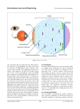
Figure 1. Structure of the cornea.
cells, suprabasal cells, and basal columnar cells located in 3.2. Keratocytes
the lower part of the epithelium. The upper three layers The keratocytes residing in the cornea are situated amidst
are comprised of differentiated surface squamous cells, the collagen fibers comprising the stoma, originating
covered with microvilli that significantly increase the from mesenchymal cells. Within this collagenous matrix,
surface of the squamous cell layer and facilitate contact keratocytes form a unified network, interconnecting
with the thin tear layer protecting the cornea. The middle with neighboring cells in a dendritic manner. These cells
layer is composed of suprabasal cells, often referred to as contain crystals and proteins that contribute to the cornea’s
Wing cells due to their wing-like shape. 13,14 Compared to transparency. Play an important role in corneal wound
squamous cells, suprabasal cells are flatter and more spread healing and scarring, keratocytes exhibit two phenotypes.
out, exhibiting elongated nuclei, with many vacuoles found One phenotype is characterized as regenerative or repair-
in proximity to the squamous cell nucleus. These cells oriented, while the other promotes cell death. During
14
divide relatively rarely, migrate toward the upper layer, and the phenotypic transformation, cells deviate from their
differentiate into squamous cells. The lowest layer of the resting state. The regenerative phenotype facilitates
epithelium is formed by basal cells in a single layer. These wound healing through fibrotic scarring and tissue repair.
cells carry out essential tasks such as replacing suprabasal However, it is noteworthy that scarring adversely affects
cells and secreting matrix factors that make up the lamina corneal transparency. 13,15
basalis and stroma. Basal cells are also responsible for the
assembly of hemidesmosomes. These functions may play a 3.3. Corneal endothelium
role in cell migration during wound healing. During eye The endothelium, constituting the cornea’s innermost
13
development, the cornea’s epithelium originates from the layer, comprises a single layer of cells located posterior to
surface epidermal ectoderm. 4 Descemet’s membrane. Comprising squamous cells that
Volume 10 Issue 2 (2024) 109 doi: 10.36922/ijb.1669