
Page 347 - IJB-10-2
P. 347
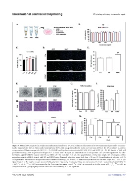
International Journal of Bioprinting                                 3D printing with drug for vascular repair


            Figure 3. NPS and NPC improve the proliferation and antioxidant effect on EPCs. (A) Schematic illustration of in vitro experimental process for curcumin-
            loaded nanoparticles (NPC), statin-loaded nanoparticles (NPS), and nanoparticles-loaded statin and curcumin (NPSC). (B) EPCs viability in various
            concentration of blank nanoparticle (NP) (N = 3). (C) Cell viability after treatment with NP, NPS, NPC, and NPSC (N = 3). (D) Results of EdU cell
            proliferation assay; 200× magnification images (N = 3). Scale bars = 100 µm. (E) Quantification of EdU-positive cells. (F) Investigation of the tube-
            formation capacity of EPCs treated with NP and NPSC (N = 3). Scale bars = 50 µm. (G) Quantification of total tube length. (H) Investigation of the
            migration capacity of EPCs treated with NP and NPSC using Transwell migration assay. Scale bars = 50 µm. (I) Quantification of migrated cell. (J)
            ROS generation was measured using fluorescence-activated cell sorting (FACS) and 2′,7′-dichlorodihydrofluorescein diacetate (H DCFDA) (N = 3). (E)
                                                                                               2
            Quantification of the reactive oxygen species (ROS) generation rates using FACS data. Values are expressed as the mean ± standard derivation (SD). *p
            < 0.05, **p < 0.01, ***p < 0.001 as compared to the Con group (untreated group);  p < 0.001 as compared to the Con group with H O ;   p < 0.001 as
                                                                                                    $$$
                                                                 ###
                                                                                                   2
                                                                                                 2
            compared to the SC group (a group treated with statin and curcumin); N.S.: not significant.
            Volume 10 Issue 2 (2024)                       339                                doi: 10.36922/ijb.1857