
Page 346 - IJB-10-2
P. 346
International Journal of Bioprinting 3D printing with drug for vascular repair
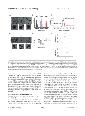
Figure 2. Characterization of statin- and curcumin-loaded nanoparticles. (A) Transmission electron microscopy (TEM) images of blank nanoparticles
(NP), statin-loaded nanoparticles (NPS), and curcumin-loaded nanoparticles (NPC). (B) Size distribution graph displaying the measured size of each
nanoparticle category. (C) Zeta potential analysis of NP, NPS, and NPC in phosphate-buffered saline (PBS; pH 7.4). (D) Storage stability study of NP, NPS,
and NPC; size distribution was analyzed after samples were incubated for 1, 3, and 7 days at 37°C. (E) Size distribution graph displaying the measured size
of each nanoparticle type after the samples were incubated for 1, 3, and 7 days at 37°C. (F) Cumulative release of statin and curcumin from nanoparticles
assessed using liquid chromatography/mass spectrometry (LC/MS) analysis.
significantly increased upon treatment with NPSC. (Figure 4B-i), the nozzles (Figure 4B-ii), and the printed
Curcumin is a type of antioxidant and can antagonize ABVs (Figure 4B-iii) are briefly described. Artificial blood
the deleterious effects of ROS. Intracellular ROS staining vessels, which can be customized in various sizes in terms
showed that the increased ROS level induced by hydrogen of their external and internal diameters, can be 3D-printed
peroxide (H O ) was suppressed in the NPSC group. To as described (Figure 4C). Twenty-four hours after the
2
2
confirm whether the nanoparticles play a role in these fabrication of ABVs, a live/dead cell staining assay was
effects, nanoparticles without being loaded with any of conducted to verify the presence of cells within the luminal
these drugs (NP) were administered (Figure 3J and K). space of the vascular construct. In the case of blood vessels
The resistance to ROS was more pronounced in the NPSC- loaded with EPCs and NPS (EPC@NPS@BV), more cells
treated group than in the group treated with statin and were observed owing to the proliferative ability of the cells
curcumin. compared with the control group (EPC@NP@BV). Blood
vessels loaded with EPCs and NPC (EPC@NPC@BV) were
3.3. 3D printing-based fabrication and found to have a morphology similar to that of the controls.
characterization of nanoparticle-loaded artificial We found that the level of cell proliferation achieved in
blood vessels EPC@NPSC@BV was similar to that observed in NPS@BV
To ensure stable transplantation of nanoparticles and (Figure 4D). The fabricated ABV was verified to contain
cells into the blood vessels, ABVs were fabricated by 3D intravascular cells within its internal structure. After we
printing (Figure 4A). The bioinks used in 3D printing confirmed the presence of a vascular framework in the
Volume 10 Issue 2 (2024) 338 doi: 10.36922/ijb.1857