
Page 431 - IJB-10-2
P. 431
International Journal of Bioprinting In vitro 3D pancreatic acinar unit
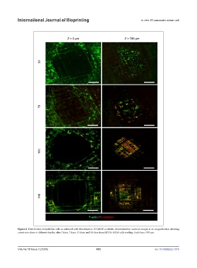
Figure 6. Distribution of epithelial cells co-cultured with fibroblasts in 3D MEW scaffolds. Representative confocal images at 4× magnification showing
constructs slices at different depths, after 3 days, 7 days, 10 days, and 14 days from HPDE-KRAS cells seeding. Scale bars: 500 µm.
Volume 10 Issue 2 (2024) 423 doi: 10.36922/ijb.1975