
Page 113 - IJB-10-3
P. 113
International Journal of Bioprinting 3D bioprinting for vascularized skin tissue engineering
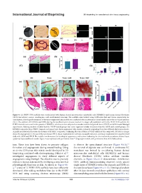
Figure 9. (A) PHBV SVN scaffolds were recellularized with human dermal microvascular endothelial cells (HDMECs) and human dermal fibroblasts
(HDFs) in indirect contact, resulting in a well-recellularized structure. The scaffolds were labeled using CellTracker Red and Green, respectively, for
visualization. Histological assessment of the tissue-engineered skin models was conducted after incubation in Green’s media and at the air–liquid interface
(ALI). The addition of HDMECs and HDFs during the recellularization process resulted in a larger cell population within the PHBV SVN scaffold. (B)
PHBV channels were used for the growth of HDMECs, which were then subjected to vascular endothelial growth factor (VEGF) or 2dDR-loaded Matrigel
experiments. Staining analysis revealed that the VEGF-loaded groups had more organized tubular structures than the 2dDR and control groups. (C)
HDMECs expanded from PHBV channels and spread onto tissue-engineered skin models, primarily originating from two different electrospun sheets.
A graph quantitatively illustrates the distance of HDMEC outgrowth, confirming that the addition of VEGF enhanced this outgrowth. (D) Macro-images
depict the angiogenesis in the case of dry eye disease (DED), tissue-engineered skin without any supplementation, and tissue-engineered skin supplemented
daily with 2dDR and VEGF. The middle row showcases the histological appearance, with arrows indicating the chorioallantoic membrane, dermal layer,
epidermal layer, and blood vessels. Figure 9 was reprinted with permission from (Copyright © 2020, with permission from ACS Publications).
141
ions. These ions have been shown to promote collagen to observe the open-channel structure (Figure 9A-b),
141
formation and angiogenesis during wound healing. Using the removal of alginate was confirmed. A continuous EC
an in vivo 3D human skin mimic model developed by 3D monolayer was formed by co-culturing human dermal
141
bioprinting combined with electrospinning, Dikici et al. microvascular endothelial cells (HDMECs) and human
investigated angiogenesis to study different aspects of dermal fibroblasts (HDFs) within artificial vascular
angiogenesis using Matrigel. The objective was to precisely channels, as Figure 9A-c-i–iii demonstrates. Anti-human
replicate a human skin model by developing a structure that CD31 antibody immunostaining observed evenly spaced
physiologically functions as skin. As shown in Figure 9A- single layers of HDMECs within the channels and HDFs on
a, a total of 12 PHBV SVN scaffolds have been efficiently the outside surfaces (Figure 9A-c-i–iii). Histological analysis
141
developed. After adding methylene blue dye to the PHBV after 14 days showed a multilayer epithelium with normal
SVN and using scanning electron microscopy (SEM) skin morphology associated with the dermis (Figure 9A-c-iv).
Volume 10 Issue 3 (2024) 105 doi: 10.36922/ijb.1727