
Page 110 - IJB-10-3
P. 110
International Journal of Bioprinting 3D bioprinting for vascularized skin tissue engineering
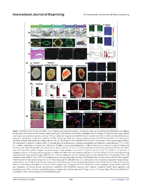
Figure 7. Development of vascularized HSEs. (A) (a) Alginate microchannels formed in 3D-printed molds, surrounded by dermal fibroblasts and collagen
gel. Keratinocytes seeded over the dermal compartment, and sacrificial layer dissolved for endothelial cell (EC) seeding. (b) Fluorescently tagged alginate
used to generate vasculature patterns; scale bar: 600 μm. Epidermal integrity and endothelial barrier function in vHSEs: (c) H&E and immunofluorescent
staining of vascularized human skin equivalent (vHSE) sections generated with induced pluripotent stem cell (iPSC)-derived ECs, for evaluating the
epidermal integrity and endothelial coating; scale bar: 250 μm. (d) Evaluation of endothelial barrier function via perfusion of fluorescently tagged dextran.
The distribution is depicted in surface plots. (e) Quantification of the fluorescence intensity, permeability, and diffusivity in microchannels. (**p < 0.005,
N = 3 HSEs). Implantation of vHSEs onto SCID mice: (f) Effect on host neovascularization. (i) Blood perfusion in Control I, Control II, vHSEs with
HUVECs, and intestinal epithelial cells (iECs) after 2 weeks; scale bar: 2.5 mm. (ii) Immunostaining of human skin equivalents (HSEs) and vHSEs
with CD31 and Ki67; scale bar: 250 μm. (iii) Quantification of host vasculature area and Ki-67-positive cells (*p < 0.05, **p < 0.005, N = 4). (g) Effect of
vasculature pattern. (i) Pictures of host vasculature in vHSEs and HSEs with microchannels; scale bars: 2.5 mm and 500 μm. (ii) Confocal image showing
perfused mouse vessels and GFP-tagged HUVECs (N = 4 for all conditions); scale bar: 500 μm. Figure 7A was reprinted from (Copyright © 2016, with
142
permission from WILEY). (B) In vitro 3D bioprinting of vascularized pigmented skin tissue. The biofabrication process involves the use of the regenHU
“BioFactoryTM 3D bio-printer system” for (a) 3D bioprinting and (b) cell cultivation. (c) 6 cm patch of pre-vascularized, pigmented human dermo-
2
epidermal skin. (d) 3D bioprinting of human melanocytes and keratinocytes follows a predetermined pattern, with cells stained and immunofluorescently
labeled. (e) Vasculature development is observed within collagen type I hydrogels via CD31 immunofluorescence imaging in human dermo-epidermal
skin substitutes. (f) Human dermal microvascular endothelial cells (HDMECs) cultured under different conditions exhibit blood capillaries and
lymphatics under CD31 and Lyve1 immunofluorescence imaging. The scale bars represent (d) 2 µm and (c, e, f) 100 µm. Figure 7B was reprinted from
129
(Copyright © 2022, with permission from SAGE Publications).
Volume 10 Issue 3 (2024) 102 doi: 10.36922/ijb.1727