
Page 188 - IJB-10-3
P. 188
International Journal of Bioprinting Bioprinted tissue-on-a-chip in drug screening
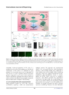
Figure 3. Schematic illustrations of different properties of bioinks. (A) The gel state during fabrication and cell culture. Reproduced with permission
from ref. (B) Chemical structure of antioxidant ink and its crosslinking process [from ref. licensed under Creative Commons Attribution (CC BY)].
78
77
(C) The printed scaffold and the viability of encapsulated cells: (Ⅰ) the printed process of the bioink, and (Ⅱ) the photographs of live/dead staining [from
.
ref. licensed under Creative Commons Attribution (CC BY)]. (D) Structure and crosslinking process of this bioink [from ref. licensed under Creative
87
85
Commons Attribution (CC BY)].
remarkable mechanical properties of PCL make it a hydrogel network. This approach may synchronously
popular choice for cardiac tissue engineering. The rebound enhance the mechanical properties and physiological
characteristic of PCL allows it to withstand the great relevance of the resulting bioinks. Each component in
pressure from ventricular systole, and the degradation of these bioinks retains its functionality without interfering
PCL lasts for 2–3 years. Synthetic polymers with lower with others. Khati et al. employed succinimidyl valerate-
87
86
gelation temperatures can be more easily modified to polyethylene glycol-succinimidyl valerate to connect
improve cell adhesion. Although these polymers with both gelatin and dECM. Gelatin enhanced the rheological
toxicity and lower cell adhesion may go against cell growth, properties, and dECM promoted the physiological
they are widely used in osteogenic manufacturing. correlation of the final bioink. The covalent linkage was
based on the reaction between the succinimidyl valerate
3.1.4. An example of the multi-component bioink group and the amino group. To ensure the stability of the
Multi-component bioinks are mixed after successive construct, the enzyme tyrosinase was used to facilitate
combinations, acquiring a stable and complementary secondary crosslinking. Moreover, the entire fabrication
Volume 10 Issue 3 (2024) 180 doi: 10.36922/ijb.1951