
Page 193 - IJB-10-3
P. 193
International Journal of Bioprinting Bioprinted tissue-on-a-chip in drug screening
factors and proteins, they cannot completely replicate 4.1. 3D-bioprinted organ-on-a-chip
drug efficiency in the human body. However, these models 3D-bioprinted organ-on-a-chip (OOC), a complex but
have the potential to substitute for animal models in the more physiologically related model, is based on microfluidic
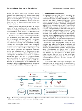
preclinical screening phase for new drugs (Figure 5). Models technology containing perfusable microfluidics, reaction
with physiological or pathological organ characteristics parts, and drug delivery channels. 3D-bioprinted OOCs,
have been used for drug metabolism analysis and drug as an advancement in the integration of 3D bioprinting
toxicity evaluation. technology with microfluidic technology, are figurative
and momentous applications of hydrogel. When combined
Disease models are broadly employed in different with bioprinting, it enables the printing of live cells in
aspects of drug screening, such as comparing drug functional hydrogel constructs that have similar spatial
efficiency underlying different mechanisms and at varying distributions to physiological microenvironments. Since
114
concentrations, as well as studying drug-drug interactions functional cells are bioprinted and perfused, the hydrogel
and determining the feasibility of combined administration. constructs could express specific factors and enzymes,
In vitro models are superior to animal experiments that possessing features of the mimicked microenvironments,
are pretty limited due to ethical issues. The technologies such as the oxygen gradient, acidity, abnormal vessels, and
in 3D-bioprinted microfluidic models complement dynamic perfusion. Once the cells have differentiated to be
each other, enabling in vitro cultures to be cultivated mature, drug solutions are applied for testing efficiency.
and stimulated artificially in miniature form accurately. Given their organ-like properties, OOCs are used
3D-bioprinted microfluidic models have successfully to explore drug toxicity for the organs by evaluating cell
115
eliminated the errors and time consumption associated with viability, metabolic activity, and biomarker expressions.
manual labor and automatically created multiple models Moreover, many studies have confirmed that liver injury
with uniform structures and components. Compared caused by hepatotoxicity from drugs is one of the factors
to the pure cell microarrays for drug screening, these contributing to the numerous challenges associated with
116
models offer more complex and bionic characteristics. 112,113 drug diagnosis and management. The renal system
3D-bioprinted microfluidic models allow for improved is also necessary for drug screening and metabolism.
interactions among cells, ECM, and relevant signaling Hepatotoxicity and nephrotoxicity slow down the drug
pathways, enabling the replication of cell behaviors and development from discovery to the clinic, increasing the
117
disease features in vivo. These models in which drug risk of failure.
reactions are observed directly can be manufactured in an Lee et al. selected PCL with low protein adsorption and
118
automated and controlled manner, reducing randomness hydrophobicity as the printed channel shells to support the
and enhancing the reliability of drug screening results. internal reaction regions. Collagen hydrogel encapsulated
Figure 5. The process of drug development from discovery to clinical experiments. The red star represents 3D-bioprinted chips discussed in this review,
which may replace animal models and 2D cultures in preclinical drug screening.
Volume 10 Issue 3 (2024) 185 doi: 10.36922/ijb.1951