
Page 194 - IJB-10-3
P. 194
International Journal of Bioprinting Bioprinted tissue-on-a-chip in drug screening
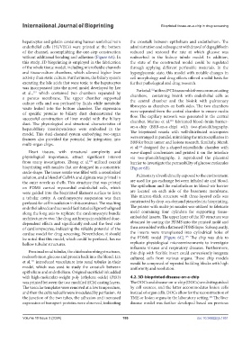
hepatocytes and gelatin containing human umbilical vein the crosstalk between epithelium and endothelium. The
endothelial cells (HUVECs) were printed at the bottom administration and subsequent withdrawal of dapagliflozin
of the channel, accomplishing the one-step construction reduced and restored the rate at which glucose was
without additional bonding and adhesion (Figure 6A). In reabsorbed in the kidney tubule model. In addition,
this study, 3D bioprinting is employed in the fabrication the state of the constructed model could be regulated
of the whole tissue model, including microfluidic channels through applying different perfusable materials. In the
and tissue-culture chambers, which allowed higher liver hyperglycemic state, this model with notable changes in
activity than static culture. Furthermore, the biliary system cell morphology and drug effects offered a solid basis for
excreting the bile acids that were toxic to the hepatocytes further pathological and drug research.
was incorporated into the novel model developed by Lee Park et al. utilized PCL to assemble three communicating
122
et al., which contained two chambers separated by chambers, containing bioink with endothelial cells as
119
a porous membrane. The upper chamber supported the central chamber and the bioink with pulmonary
culture cells and was perfused by fluids while metabolic
waste leaked into the bottom chamber. The expression fibrocytes as chambers on both sides. The two chambers
of specific proteins to biliary duct demonstrated the were separated from the central chamber to ensure media
successful construction of liver model with the biliary flow. The capillary network was generated in the central
123
duct. The physiological and structural characteristics of chamber. Marino et al. fabricated blood–brain barrier-
on-a-chip (BBB-on-a-chip) with two-photolithography.
hepatobiliary interdependence were embodied in the
model. This dual-channel system embodying two-organ The bioprinted vessels with well-distributed micropores
features also provided the potential for integration into were arranged in parallel, mimicking the microcapillaries in
multi-organ chips. BBB for brain tumor and lesions research. Similarly, Mandt
et al. designed the x-shaped microfluidic chamber with
124
Heart tissues, with structural complexity and wave-shaped condensates and printed it on the interface
physiological importance, attract significant interest via two-photolithography. It reproduced the placental
from many investigators. Zhang et al. utilized coaxial barrier to investigate the permeability of glucose molecules
120
bioprinting with nozzles that are designed in a concentric (Figure 6B).
circle shape. The inner nozzle was filled with a crosslinked
solution, and a blend of GelMA and alginate was printed in Pulmonary alveoli directly exposed to the environment
the outer nozzle as shells. This structure that was printed are used for gas exchange between inhaled air and blood.
on PDMS carried myocardial endothelial cells, which The epithelium and the endothelium in blood–air barrier
were guided into the bioprinted filament surface to form are located on each side of the basement membrane.
a tubular cavity. A cardiomyocyte suspension was then This micron-thick structure with three-layered cells was
perfused for cell inoculation in this construct. The resulting constructed by drop-on-demand piezoelectric bioprinting.
endothelialized cardiac model facilitated cell growth aligned The printer with multi-jet nozzles was utilized to fabricate
along the long axis to replicate the cardiomyocyte bundle mold containing four cylinders for supporting tissue-
architecture in vivo. The drug azithromycin exhibited dose- embedded inserts. The upper layer of the 3D structure was
dependent effects and significantly reduced the beat rate obtained by casting the PDMS into the printed mold and
of cardiomyocytes, indicating the reliable potential of the then assembled with a flattened PDMS layer. Subsequently,
cardiac model for drug screening. Nevertheless, it should the inserts were transplanted into cylindrical holes of
125
be noted that this model, which could be perfused, has no the PDMS model (Figure 6C). The chip was able to
hollow tubular structures. replicate physiological microenvironments to investigate
influenza viruses and respiratory diseases. Furthermore,
Proximal renal tubules, the chief reabsorbing structures, this chip with flexible insert could conveniently integrate
reabsorb most glucose and protein back into the blood. Lin cultured cells from various organs. These chip models
121
et al. introduced vasculature into renal tubules in their would be composed of repeated building blocks with high
model, which was used to study the crosstalk between uniformity and resolution.
epithelium and endothelium. Original sacrificial ink added
with high-molecular-weight poly (ethylene oxide) (PEO) 4.2. 3D-bioprinted disease-on-a-chip
was printed between the two modified ECM casting layers. The OOCs and disease-on-a-chip (DOCs) are distinguished
The vascular templates were removed at a low temperature, by cell sources, and the latter accommodates lesion cells
and then the cultured cells were inoculated by perfusion. At instead of organ cells. DOCs allow for the reconstruction of
the junction of the two tubes, the adhesion and increased TME or lesion organs in the laboratory setting. The liver
126
expression of transport proteins were observed, indicating disease model was further developed based on previous
Volume 10 Issue 3 (2024) 186 doi: 10.36922/ijb.1951