
Page 195 - IJB-10-3
P. 195
International Journal of Bioprinting Bioprinted tissue-on-a-chip in drug screening
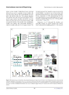
studies on liver models. Gelatin-based bioink dissolved structure possessed two channels compartmentalized by
afterward was used to encapsulate activated stellate cells printed bioinks. It was known that activated stellate cells
that induced liver fibrosis. The human HepaRG cells were generated excessive ECM due to the increased expression
laid in the dECM of the liver. Two adjacent chambers of key components in ECM, demonstrating that this
were printed with poly-(ethylene/vinyl acetate) (PEVA), pathological model was successfully fabricated and could
and the upper chamber was filled with dECM. Secondly, be used to evaluate drug efficiency for inhibiting stellate
127
PEVA was added again on the first-printed chamber walls cell activation (Figure 6D).
to deepen the volume of the chamber. After consolidation, Nothdurfter et al. fabricated the chamber and
128
bioinks were printed and subsequently dissolved in perfusable channels on PMMA through laser etching,
order to inoculate cells and form the upper channel. The which addressed the shedding problem of the vascular
Figure 6. Fabrication processes of microfluidic chips for mimicking disease or organ microenvironments. (A) Cell-bioprinted liver-on-a-chip in one-step
fabrication (reproduced with permission from ref. ). (B) Placental barrier-on-a-chip under two-photon polymerization (reproduced with permission
118
from ref. ). (C) Inkjet-bioprinting lung-on-a-chip(Reproduced with permission from ref. ).(D) 3D liver fibrosis-on-a-chip (reproduced with permission
125
124
from ref. ). (E) Micro-vascularized neuroblastoma-on-a-chip in medium throughput (from ref. licensed under Creative Commons Attribution 4.0
128
127
license).
Volume 10 Issue 3 (2024) 187 doi: 10.36922/ijb.1951