
Page 462 - IJB-10-3
P. 462
International Journal of Bioprinting Biomimetic scaffolds for tendon healing
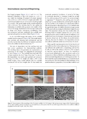
the ImageJ program (Figure 4A-i–iii and A-iv–vi). The previously mentioned conditions. A period of 85 days,
intended dimensions of the CAD design are 1 cm × 1 as previously described in literature, would be enough
cm, while the resulting 3D-printed structures measure for the cells to produce ECM and for the tendon to begin
1.02 ± 0.03 cm × 0.99 ± 0.02 cm in length and width. The to regenerate. 59,60 Meanwhile, the swelling ratio for each
CAD design’s height was set at two layers (resulting height sampling time was calculated by correlating the weight of
of 1.44 mm). The actual height of the printed scaffolds is the wet scaffold to the weight of the dehydrated scaffold.
1.49 ± 0.16 mm. This indicates that the ink enables the The data presented in the graph (Figure 5A-ii) indicate that
fabrication of scaffolds with a geometry very similar to that during the first hour, the scaffold absorbed a large amount
of the CAD design. Furthermore, the structure maintained of PBS, practically increasing its dehydrated weight by nine
its shape even before undergoing crosslinking. Once times. The maximum selling ratio was reached after 3 h,
the parameters had been optimized, the scaffolds were following which it remains constant for up to 24 h. This
automatically printed in culture dishes (Figure 4A-vii). increase in water content could decrease the stiffness of the
In the cryo-SEM images, the surface of the scaffolds scaffold and favor the transport of nutrients and molecules
exhibits a porous structure (Figure 4B). This rough surface of interest (important for the release of growth factors).
may promote cell adhesion and proliferation, and the Likewise, they may be related to the degradation process
porosity may facilitate the diffusion of nutrients and other the scaffold underwent in the first several days of culture.
molecules within the microenvironment. 57,58 Other equally important parameters for the function of
The rate of degradation and the swelling ratio are the scaffold are the mechanical properties. These properties
two crucial parameters for characterizing scaffolds. can influence the proliferation, morphology, and activity
The resulting values of degradation are represented as a of cells, as well as the release of factors or the transport
percentage of the weight loss (degradation percentage) of nutrients, among others. In turn, the mechanical
61
vs. time (Figure 5A-i). The scaffolds lose practically 30% properties are influenced by other parameters such as the
of their weight in the first 7 days. From day 7 to day 42, composition of the scaffold, the crosslinking process, or the
the degradation was much more gradual and sustained, aforementioned degradation and swelling parameters. To
losing only 10% of the weight (in total, a 40% of the determine the mechanical performance, a compression test
initial weight). These results indicate that the scaffolds was performed. This test enabled the determination of the
maintained 60% of their weight after 85 days under the initial mechanical properties of the acellular scaffolds and
Figure 4. Characterization of the morphology of the 3D-printed scaffolds. (A) CAD designs (i–iii) and macroscopic images of the 3D-printed scaffolds
(iv–vi). These scaffolds could also be printed in an automated way on culture plates (vii). (B) Cryo-SEM images of the surface of the scaffolds at different
magnifications (from left to right: 120x, 300x, 1000x).
Volume 10 Issue 3 (2024) 454 doi: 10.36922/ijb.2632