
Page 459 - IJB-10-4
P. 459
International Journal of Bioprinting Biomechanical mimic-based artificial oviduct system
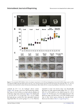
Figure 3. Two pronuclear (PN) fertilized embryo development speed ratios on the 3D-bioprinted gelatin methacryloyl (GelMA) culture system. (A) The
3D-bioprinted GelMA hydrogel culture system. Scale bar: 5 mm. (B) Development speed ratios of two PN embryos into blastocytes (BLs). *p < 0.05. (C)
Assessment of preimplantation embryo development in the hydrogel culture system with the corresponding timeframe. Abbreviations: Con, Control.
cultured for 96 h on the hydrogel culture system required to reach the morula stage was dramatically
(Figure 3B). Using a real-time cell monitoring system, decreased in both experimental groups (8%: 57 ± 33 h;
the times at which embryos reached each developmental 10%: 53 ± 38 h) relative to the control group (62 ± 93 h)
embryonic stage were measured (Figure 3B). Blastocysts (Figure 3B). Furthermore, the developmental rate to the
in the 8% (65 ± 7.3 h) and 10% (70 ± 3.8 h) GelMA blastocyst stage was 20% faster in the 10% GelMA group
hydrogel groups reached the fully expanded blastocyst than in the control group during the morula-to-late-
stage faster than the control group (73 ± 5.1 h). The time blastocyst transition (Table S1, Supporting File).
Volume 10 Issue 4 (2024) 451 doi: 10.36922/ijb.3346