
Page 579 - IJB-10-4
P. 579
International Journal of Bioprinting 3D-printing silicone patient-specific soft-tissue expander
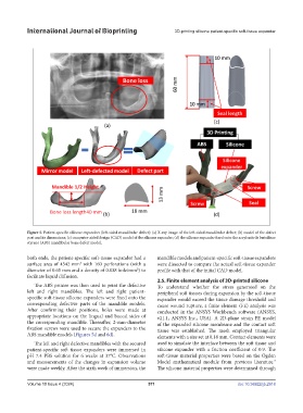
Figure 5. Patient-specific silicone expanders (left-sided mandibular defect): (a) X-ray image of the left-sided mandibular defect; (b) model of the defect
part and its dimensions; (c) computer-aided design (CAD) model of the silicone expander; (d) the silicone expander fixed onto the acrylonitrile butadiene
styrene (ABS) mandibular bone defect model.
both ends, the patient-specific soft-tissue expander had a mandible models and patient-specific soft-tissue expanders
3
surface area of 4342 mm with 160 perforations (with a were dissected to compare the actual soft-tissue expander
2
diameter of 0.65 mm and a density of 0.038 hole/mm ) to profile with that of the initial CAD model.
facilitate liquid diffusion.
2.5. Finite element analysis of 3D-printed silicone
The ABS printer was then used to print the defective To understand whether the stress generated on the
left and right mandibles. The left and right patient- peripheral soft tissues during expansion by the soft-tissue
specific soft-tissue silicone expanders were fixed onto the expander would exceed the tissue damage threshold and
corresponding defective parts of the mandible models. cause wound rupture, a finite element (FE) analysis was
After confirming their positions, holes were made at conducted in the ANSYS Workbench software (ANSYS,
appropriate locations on the lingual and buccal sides of v21.1, ANSYS Inc., USA). A 2D-plane strain FE model
the corresponding mandible. Thereafter, 2-mm-diameter of the expanded silicone membrane and the contact soft
fixation screws were used to secure the expanders to the tissue was established. The mesh employed triangular
ABS mandible models (Figures 5d and 6d). elements with a size set at 0.18 mm. Contact elements were
The left and right defective mandibles with the secured used to simulate the interface between the soft tissue and
patient-specific soft tissue expanders were immersed in silicone expander with a friction coefficient of 0.9. The
pH 7.4 PBS solution for 6 weeks at 37°C. Observations soft-tissue material properties were based on the Ogden
and measurements of the changes in expansion volume Model mathematical module from previous literature.
7
were made weekly. After the sixth week of immersion, the The silicone material properties were determined through
Volume 10 Issue 4 (2024) 571 doi: 10.36922/ijb.2918