
Page 25 - IJB-5-2
P. 25
Mishbak, et al.
and different reaction times. The results show that both of functionalized alginate polymers obtained after 8 and
the storage and loss modulus are higher in samples 24 h of reaction time, mixed with 0.5-1.5% w/v, and of
obtained after high functionalization times. In both cases, VA-086 photoinitiator solutions.
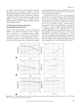
the complex modulus increases with frequency, while the As observed from Figures 12 and 13 by increasing the
variation of Gʹ and Gʺ with strain shows a shift point after curing time Gʹ increases becoming significantly higher
which Gʺ becomes higher than Gʹ. This shift occurs at than Gʺ. It is also possible to observe that by increasing
high frequencies for alginate samples obtained with high the photoinitiator concentration the gelation time, which
functionalization times. corresponds to the time point where the Gʹ curve crosses
the Gʺ curve, decreases. Notably observed that by
3.3 Rheological Changes during the increasing the amount of photoinitiators concentration
Photopolymerization Process Gʹ seems to tend to a plateau which corresponds to
a verification stage of the curing process [35,36] . The
Based on the characterization of the pre-polymerized possible explanation for this observation is that, for the
samples, only systems containing 2% w/v of alginate layer thickness considered in this study (~100 µm), the
were considered for photopolymerization studies. photoinitiator concentration is approaching a critical
The curing kinetics was assessed by monitoring the value. By increasing the photoinitiator concentration
variation of Gʹ and Gʺ at room temperature through a above the critical values, the polymerization occurs very
controlled frequency of (1Hz). Photorheology was used fast at the polymer surface, reducing the light penetration
to characterize the curing process (photopolymerization) and, consequently, the overall polymerization reduces.
a
b
c
Figure 14. 2% wt. methacrylate alginate hydrogel with different concentration of VA-086 functionalized for 8 h. (a) 0.5 w/v % of VA-086,
(b) 1 w/v % of VA-086, (c) 1.5 w/v % of VA-086.
International Journal of Bioprinting (2019)–Volume 5, Issue 2 21