
Page 402 - IJB-10-5
P. 402
International Journal of Bioprinting DEX-Loaded PLGA microspheres enhance cartilage regeneration
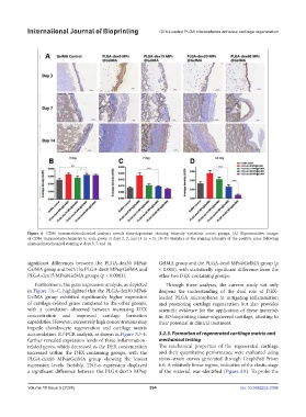
Figure 6. CD86 immunohistochemical analysis reveals time-dependent staining intensity variations across groups. (A) Representative images
of CD86 immunohistochemistry in each group at days 3, 7, and 14 (n = 5). (B–D) Statistics of the staining intensity of the positive areas following
immunohistochemical staining at days 3, 7, and 14.
significant differences between the PLGA-dex30 MPs@ GelMA group and the PLGA-dex0 MPs@GelMA group (p
GelMA group and both the PLGA-dex0 MPs@GelMA and < 0.001), with statistically significant difference from the
PLGA-dex15 MPs@GelMA groups (p < 0.0001). other two DEX-containing groups.
Furthermore, the gene expression analysis, as depicted Through these analyses, the current study not only
in Figure 7A–C, highlighted that the PLGA-dex30 MPs@ deepens the understanding of the dual role of DEX-
GelMA group exhibited significantly higher expression loaded PLGA microspheres in mitigating inflammation
of cartilage-related genes compared to the other groups, and promoting cartilage regeneration but also provides
with a correlation observed between increasing DEX scientific evidence for the application of these materials
concentration and improved cartilage formation in 3D-bioprinting tissue-engineered cartilage, attesting to
capabilities. However, excessively high concentrations may their potential in clinical treatment.
impede chondrocyte regeneration and cartilage matrix
accumulation. RT-PCR analysis, as shown in Figure 7D–F, 3.2.3. Formation of regenerated cartilage matrix and
further revealed expression levels of three inflammation- mechanical testing
related genes, which decreased as the DEX concentration The mechanical properties of the regenerated cartilage
increased within the DEX-containing groups, with the and their quantitative performance were evaluated using
PLGA-dex60 MPs@GelMA group showing the lowest stress–strain curves generated through GraphPad Prism
expression levels. Notably, TNF-α expression displayed 6.0. A relatively linear region, indicative of the elastic stage
a significant difference between the PLGA-dex15 MPs@ of the material, was identified (Figure 8A). To probe the
Volume 10 Issue 5 (2024) 394 doi: 10.36922/ijb.3396