
Page 399 - IJB-10-5
P. 399
International Journal of Bioprinting DEX-Loaded PLGA microspheres enhance cartilage regeneration
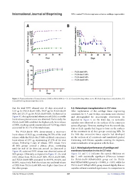
Figure 3. Pharmacokinetics of dexamethasone (DEX) in PLGA MPs (n = 3). (A) Cumulative drug release curve. (B) Daily drug release concentration. (C)
Cumulative drug release proportion curve.
that the total DEX released over 45 days amounted to 3.2. Heterotopic transplantation in C57 mice
5.23 µg for PLGA-dex15 MPs, 59.07 µg for PLGA-dex30 After implantation of the cartilage tissue engineering
MPs, and 251.55 µg for PLGA-dex60 MPs. As depicted in constructs for 3, 7, and 14 days, specimens were obtained
Figure 3C, during the initial release period (24 h), a notable and photographed for macroscopic observation. As
burst release phenomenon was observed. Particularly, the depicted in Figure 4, on the third day, no noticeable
PLGA-dex60 MPs exhibited the highest early burst release capsules were observed on the surfaces of the constructs
of DEX, reaching a peak concentration of 74.056 µg, which across all groups. However, starting from the seventh day,
accounted for 26.17% of the total release. thin or thick capsules had begun to form on the surfaces
The PLGA-dex30 MPs demonstrated a maximum of the constructs in all four groups containing MPs. By
burst release of 38.22 μg, constituting 54.75% of the total the 14th day, connective tissue capsules had developed
release, while the PLGA-dex15 MPs exhibited a maximum on the surfaces of all constructs and manifested gradual
burst release of 2.97 μg, representing 47.17% of the total thickening, with fibrous capsules covering the implants,
release. Following 3 days of release, DEX release from which is indicative of integration with the host.
all MPs groups entered a plateau phase, continuing
until the end of the detection period. As illustrated in 3.2.1. Histological performance of cartilage and
Figure 3B, sustained DEX release was observed across all membrane formation in vivo in C57 mice
MPs groups. Ultimately, as depicted in Figure 3C, the total Figure 5A and C–E indicates the capsule thickness on
DEX release from PLGA-dex15 MPs, PLGA-dex30 MPs, day 7, with statistically significant differences between
and PLGA-dex60 MPs amounted to 83.07%, 84.62%, and the PLGA-dex30 MPs@GelMA group and the PLGA-
88.89%, respectively. Both the release rate and total release dex0 MPs@GelMA group (p < 0.0001, p < 0.05), while the
of DEX from PLGA-dex60 MPs were higher than those of PLGA-dex15 MPs@GelMA group showed slightly thinner
the other groups. capsules without statistical significance. In comparison to
Volume 10 Issue 5 (2024) 391 doi: 10.36922/ijb.3396