
Page 17 - IJB-10-6
P. 17
International Journal of Bioprinting 3D-bioprinted multicellular lung organoids
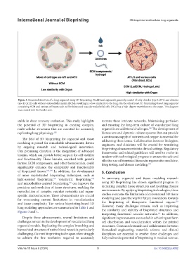
Figure 3. Improved structure of a lung organoid using 3D bioprinting. Traditional organoids generally consist of only alveolar type I (AT1) and alveolar
type II (AT2) cells without extracellular matrix (ECM), resulting in a low similarity to the lung. On the other hand, 3D bioprinting-based lung organoid
containing ECM and various cell types such as fibroblasts and vascular endothelial cells (ECs) has a high-degree resemblance to the organ. This diagram
was created with BioRender.com.
stable in shear recovery evaluation. This study highlights recreate these intricate networks. Maintaining perfusion
the potential of 3D bioprinting in creating complex, and ensuring the long-term culture of vascularized lung
multi-cellular structures that are essential for accurately organoids are additional challenges. The development of
108
replicating lung physiology. 102 bioreactors and dynamic culture systems that can provide
a continuous supply of nutrients and oxygen is essential for
The field of 3D bioprinting for organoid and tissue addressing these issues. Collaboration between biologists,
modeling is poised for remarkable advancements, driven engineers, and clinicians will be crucial for translating
by ongoing research and technological innovation. bioprinting advancements into clinical settings. Regulatory
One promising direction is the integration of advanced frameworks and ethical guidelines will need to evolve in
bioinks, which can provide better support for cell viability tandem with technological progress to ensure the safe and
and functionality. These bioinks, enriched with growth effective use of bioprinted tissues in regenerative medicine,
factors, ECM components, and other biomolecules, could drug testing, and disease modeling. 109
significantly enhance the complexity and functionality
of bioprinted tissues. 103,104 In addition, the development 5. Conclusion
of more sophisticated bioprinting techniques, such as
light-assisted bioprinting, volumetric bioprinting, In summary, organoid and tissue modeling research
105
106
and microfluidics-assisted bioprinting, can improve the using 3D bioprinting has shown significant progress in
107
precision and resolution of tissue structures, enabling the recreating complex tissue structures and modeling disease
reproduction of complex vascular networks and organ- environments. By applying bioprinting technologies, these
specific microstructures. These advancements are crucial studies overcome the limitations of conventional 3D tissue
for overcoming current limitations in vascularization modeling and pave the way for future innovations towards
110
and tissue complexity. The various bioprinting-based 3D the bioprinting of therapeutic functional organs.
lung modeling approaches are summarized in Table 1 and However, many challenges remain, such as improving
Figures 3 and 4. the similarity and stability of bioprinted structures and
integrating functional vascular networks. In addition,
111
Despite these advancements, several limitations and significant improvements are needed in achieving uniform
challenges remain in the development of vascularized lung cell distribution and vascularization within the printed
organoid models. Replicating the complex branching and structures. Continued research and collaboration between
hierarchical structure of native blood vessels is particularly biomedical engineering, materials science, and clinical
challenging. Current bioprinting techniques often struggle disciplines are essential to resolve these challenges and
to achieve the fine resolution required to accurately fully realize the potential of bioprinting in medical science.
Volume 10 Issue 6 (2024) 9 doi: 10.36922/ijb.4092