
Page 143 - IJB-7-1
P. 143
Rodriguez-Salvador, et al.
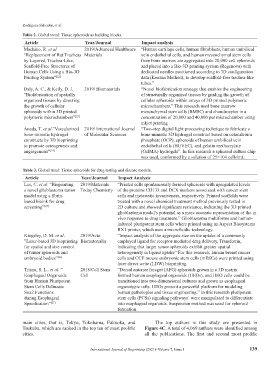
Table 2. Global trend: Tissue spheroids as building blocks.
Article Year/Journal Impact analysis
Machino, R. et al 2019/Advanced Healthcare “Human cartilage cells, human fibroblasts, human umbilical
“Replacement of Rat Tracheas Materials vein endothelial cells, and human mesenchymal stem cells
by Layered, Trachea-Like, from bone marrow are aggregated into 20,000 cell spheroids
Scaffold-Free Structures of and placed into a Bio-3D printing system (Regenova) with
Human Cells Using a Bio-3D dedicated needles positioned according to 3D configuration
Printing System” [22] data (Kenzan Method), to develop scaffold-free trachea-like
tubes.”
Daly, A. C., & Kelly, D. J. 2019/ Biomaterials “Novel biofabrication strategy that enables the engineering
“Biofabrication of spatially of structurally organized tissues by guiding the growth of
organised tissues by directing cellular spheroids within arrays of 3D printed polymeric
the growth of cellular microchambers.” This research used bone marrow
spheroids within 3D printed mesenchymal stem cells (BMSC) and chondrocytes in a
polymeric microchambers” [23] concentration of 20,000 and 40,000 per microchamber using
inkjet printing
Anada, T. et al ”Vascularized 2019/ International Journal “Two-step digital light processing technique to fabricate a
bone-mimetic hydrogel of Molecular Sciences bone-mimetic 3D hydrogel construct based on octacalcium
constructs by 3D bioprinting phosphate (OCP), spheroids of human umbilical vein
to promote osteogenesis and endothelial cells (HUVEC), and gelatin methacrylate
angiogenesis” [24] (GelMA) hydrogels”. In this research a spheroid culture chip
was used, conformed by a solution of 25×104 cells/mL
Table 3. Global trend: Tissue spheroids for drug testing and disease models.
Article Year/Journal Impact Analysis
Lee, C. et al. “Bioprinting 2019/Materials “Printed cells spontaneously formed spheroids with upregulated levels
a novel glioblastoma tumor Today Chemistry of the proteins CD133 and DCX markers associated with cancer stem
model using a fibrin- cells and metastatic invasiveness, respectively. Printed scaffolds were
based bioink for drug treated with a novel chemical treatment method previously tested in
screening” [25]. 2D culture and showed significant resistance, indicating the 3D printed
glioblastoma model’s potential as a more accurate representation of the in
vivo response to drug treatment.” Glioblastoma multiforme and human-
induced pluripotent stem cells where printed using an Aspect Biosystems
RX1 printer, which uses a microfluidic technology.
Kingsley, D. M. et al. 2019/Acta “Impact analysis of the aggregate size on the uptake of a commonly
”Laser-based 3D bioprinting Biomaterialia employed ligand for receptor-mediated drug delivery, Transferrin,
for spatial and size control indicating that larger tumor spheroids exhibit greater spatial
of tumor spheroids and heterogeneity in ligand uptake” For this research, human breast cancer
embryoid bodies” [26] cells and CCE mouse embryonic stem cells (mESCs) were printed using
laser direct write (LDW) bioprinting.
Trisno, S. L.. et al. ” 2018/Cell Stem “Dorsal anterior foregut (AFG) spheroids grown in a 3D matrix
Esophageal Organoids Cell formed human esophageal organoids (HEOs), and HEO cells could be
from Human Pluripotent transitioned into two-dimensional cultures and grown as esophageal
Stem Cells Delineate organotypic rafts. HEOs present a powerful platform for modeling
Sox2 Functions human pathologies and tissue engineering.” In this research pluripotent
during Esophageal stem cells (PCSs) signaling pathways´ were manipulated to differentiate
Specification” [27] into esophageal organoids. Suspension method was used for spheroid
formation.
main cities, that is, Tokyo, Yokohama, Fukuoka, and The top authors in this study are presented in
Tsukuba, which are ranked in the top ten of most prolific Figure 4C. A total of 4,069 authors were identified among
cities. all the publications. The first and second most prolific
International Journal of Bioprinting (2021)–Volume 7, Issue 1 139