
Page 145 - IJB-7-1
P. 145
Rodriguez-Salvador, et al.
Table 5. Global trend: Complementary studies.
Article Year/Journal Impact Analysis
Nakagawa, K., & Kishimoto, T. 2019/Biotechniques “Unlabeled optical metabolic imaging of cultured living
“Unlabeled image analysis-based cells. This imaging technique is based on motion vector
cell viability assay with intracellular analysis with a block-matching algorithm to compare
movement monitoring” [31] sequential time-lapse images. Motion vector analysis
evaluates the movement of intracellular granules observed
with a phase-contrast microscope. This assay can measure
cellular viability at a single-cell level without requiring
any reagents”. In this research, human osteosarcoma
U2OS cells, human colon carcinoma Caco-2 cells and
human hepatoma HepG2 cells were used.
Wu, H. et al. “Electrical impedance 2018/Analyst “In silico and in vitro cell viability inside large cell
tomography for real-time and label-free spheroids can be monitored in real time and label-free
cellular viability assays of 3D tumour with electrical impedance tomography (EIT). The results
spheroids” [32] show the potential of EIT for non-destructive real-time
and label-free cellular assays in the miniature sensor,
providing physiological information in the applications
of the 3D drug screening and tissue engineering.” MCF-7
breast cancer cells were used, and the liquid overlay
technique was adopted to form cells spheroids on the
hydrogel surface. Cell suspension with 1×10⁴ cells were
seeded onto each microplate well.
Parrish, J et al. “A 96-well microplate 2018/Lab on a Chip “Platform to address the experimental and in vivo disparity
bioreactor platform supporting in throughput and both system complexity (by supporting
individual dual perfusion and high- multiple in situ assessment methods) and tissue complexity
throughput assessment of simple or (by adopting a construct-agnostic format). It describes the
biofabricated 3D tissue models” [33] potential of a scalable dual perfusion bioreactor platform
for parenchymal and barrier tissue constructs to support
a broad range of multi-organ-in-a-chip applications”.
In this research human umbilical cord-derived vascular
endothelial cells (HUVEC), bone marrow-derived
mesenchymal stromal cells (MSC), human ovarian cancer
cells and human foreskin-derived fibroblast were used.
is the most prolific journal with 62 documents, followed
by Acta Biomaterialia which has almost half the number
of articles with 36. Tissue Engineering – Part A claims
the third place with 32 publications. These three journals
comprise 52% of all the documents in the top ten list.
These journals focus on either biomaterial structure,
function, and clinical application or in therapeutic
strategies to regenerate tissue – the topics closely related
to tissue spheroids.
Furthermore, the institutions with the highest
numbers of publications were also identified, as shown
in Figure 4E. Overall, 840 institutions were identified
worldwide but the most prolific institutions are directly
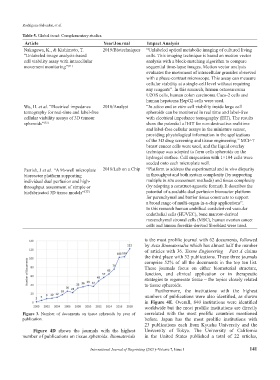
Figure 3. Number of documents on tissue spheroids by year of correlated with the most prolific countries mentioned
publication. before. Japan has the most prolific institutions with
23 publications each from Kyushu University and the
Figure 4D shows the journals with the highest University of Tokyo. The University of California
number of publications on tissue spheroids. Biomaterials in the United States published a total of 22 articles,
International Journal of Bioprinting (2021)–Volume 7, Issue 1 141