
Page 247 - IJB-8-3
P. 247
Li, et al.
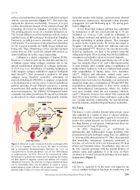
authors claimed that they have printed a whole living heart ventricles printed with human cardiomyocytes showed
with the vascular networks (Figure 3) . This report has synchronized contractions, directional action potential
[82]
captured the attention worldwidely. However, it is just propagation, and wall thickening up to 14% during peak
another development branch of the extrusion-based RP systole (Figure 4).
technology developed in Tsinghua University in 2003. It was found that collagen gelation was controlled
The printing process occurs in a medium instead of air. by modulation of pH and could provide up to 10 μm
The obvious differences of this technique with the former resolution on printing. Cells could be embedded in
reported series of RP techniques developed in Tsinghua the collagen hydrogel and introduced into the scaffold
University include polymeric materials, cell types, and through embedding of gelatin spheres. This technique
CAD models. The movement of blood has been mimicked is similar to the double-nozzle 3D printer developed in
but the vascular networks are totally forged without any Tsinghua University, in which two different materials
living cells. Thus, 3D printing of thick vascularized heart have been printed [3,43,47] . However, the vascular networks,
tissues that can fully match the patient still remains an including capillaries, are hard to be printed using the
unmet challenge in cardiac organ engineering. collagen hydrogels. There is a long way for the multiscale
Collagen is a major component of human ECMs. vasculature and trileaflet valves to be used clinically.
However, it is hard to replicate the structure and function Many other 3D printing heart tissues are still on the
of human organs using collagen solutions due to the way. For example, Duan et al. used a flat-shaped model
special solidification properties of collagen molecules. to print trileaflet valve conduits using a combination of
In 2019, Lee et al. described a 3D printing technique to methacrylated hyaluronic acid and methacrylated gelatin
build complex collagen scaffolds to engineer biological with encapsulation of human aortic valve interstitial
heart tissues . They presented a method to 3D print cells . Stiffness and adhesivity control aortic valve
[83]
[84]
collagen using freeform reversible embedding of interstitial cell behavior within hyaluronic acid-based
suspended hydrogels (FRESHs) to engineer components hydrogels have been achieved. Hockaday et al. used axially
of the human heart at various scales. Control of pH-driven symmetric shape and a combination of 700 and 8000 Mw
gelation provides 20 μm filament resolution, a porous poly(ethylene glycol)diacrylate to print valve conduits
microstructure that enables rapid cellular infiltration and with biomechanical heterogeneity, where the leaflets
microvascularization. The FRESH 3D-bioprinted hearts were more flexible, while the root remained relatively
accurately reproduce patient-specific anatomical structure rigid . However, evidence has shown that vascularized
[85]
as determined by micro-computed tomography. Cardiac heart 3D printing still faces many challenges, such as the
real vascular network construction, the anti-thrombotic
material selection, and physiological function realization.
4.2. Lung
The lung is a very complex internal immunologic organ
who responds in a variety of ways to inhaled antigens,
infectious materials, or saprophytic agents. It’s commonly
known that certain diseases are linked with occupations
like lung disease in coal miners. Until present, there are
few references for lung 3D printing.
In 2019, Grigoryan et al. established an
intravascular and multivascular design freedoms with
photopolymerizable hydrogels using food dye additives
as biocompatible photoabsorbers . They demonstrated
[86]
that monolithic transparent hydrogels, produced in
Figure 3. Omentum tissue is extracted from the patient and while minutes, can efficiently mimic the intravascular 3D fluid
the cells are separated from the matrix, the latter is processed mixers and bicuspid valves. Nevertheless, they are not
into a personalized thermoresponsive hydrogel, the cells are the true vascular networks with the typical features of
reprogrammed to become pluripotent and are then differentiated arteries, veins, and capillaries. It is far away for these
to cardiomyocytes and ECs, followed by encapsulation within constructs to be used as bioartificial lungs for anti-suture
the hydrogel to generate the bioinks used for printing, the bioinks implantation (Figure 5).
are then printed to engineer vascularized patches and complex
cellularized structures, the resulting autologous engineered tissue 4.3. Liver
can be transplanted back into the patient, to repair or replace
injured/diseased organs with low risk of rejection. (from ref. [82] The liver is regarded to be the most vital organ for its
licensed under Creative Commons Attribution license). critical multiple biological functions in metabolism and
International Journal of Bioprinting (2022)–Volume 8, Issue 3 239